Copyright
©The Author(s) 2025.
World J Clin Cases. May 26, 2025; 13(15): 102691
Published online May 26, 2025. doi: 10.12998/wjcc.v13.i15.102691
Published online May 26, 2025. doi: 10.12998/wjcc.v13.i15.102691
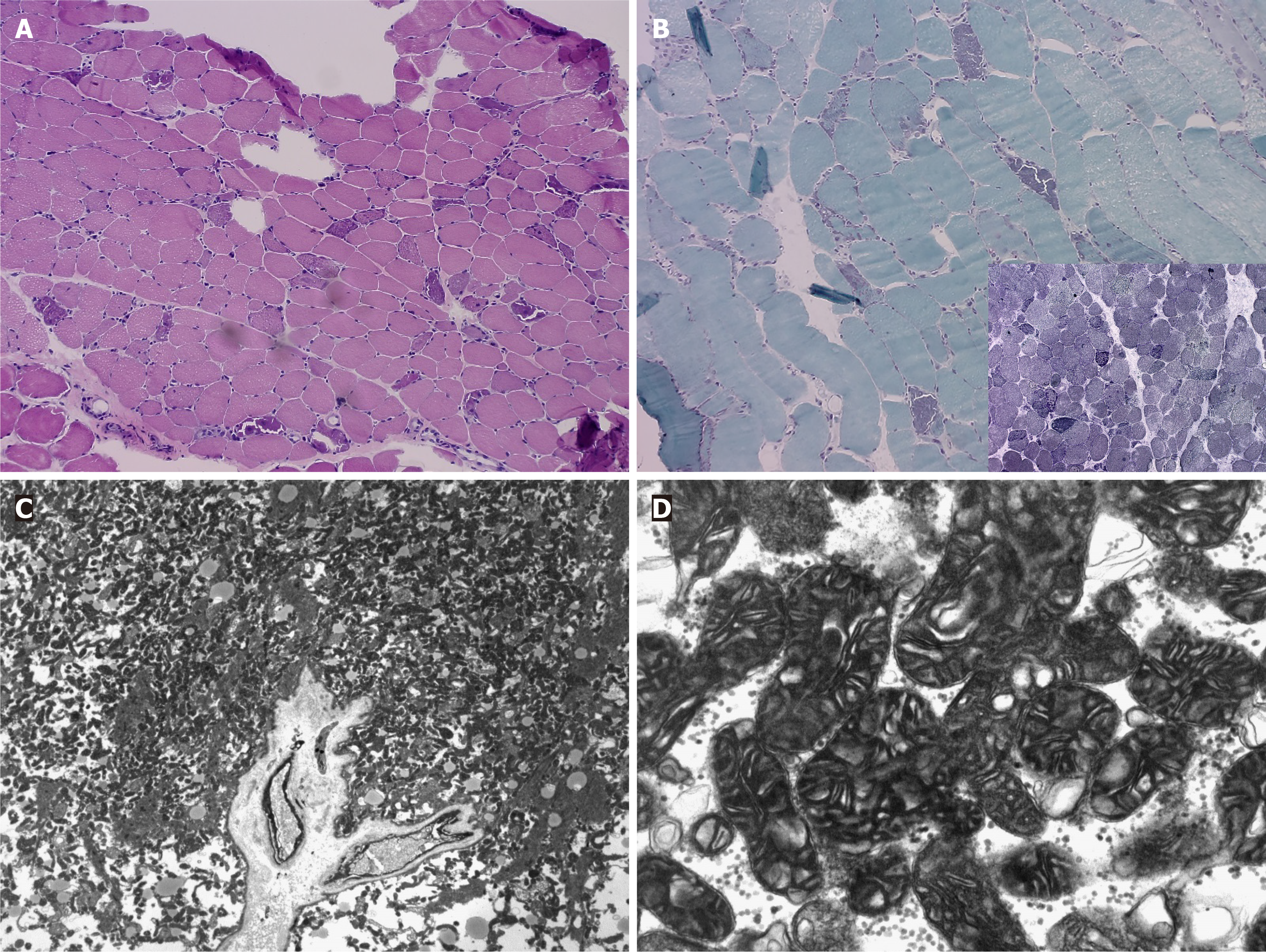
Figure 1 Microscopic findings of the biceps bracii muscle.
A: Moderate variation in myofiber size and many degenerating and regenerating myofibers are shown. Circular/Spherical atrophic myofibers are frequently observed (hematoxylin and eosin, × 100); B: On modified Gomori-trichrome stain, ragged red fibers with increased subsarcolemmal and intermyofibrillar red staining are frequently seen (modified Gomori-trichrome, × 200). (Inlet) On succinate dehydrogenase (SDH) staining, strong positivity is present in many myofibers (SDH, × 200); C: Subsarcolemmal accumulation of swollen mitochondria is frequently noted in electron microscope (EM) (EM, × 2000); D: Many abnormally enlarged mitochondria are noted (EM, × 30000).
- Citation: Park SY, Hong SM, Lee HY, Kim MY, Lee HK, Han JY, Cho HJ, Oh SI, Lee H. Mitochondrial myopathy revealed postoperative acute respiratory failure: A case report. World J Clin Cases 2025; 13(15): 102691
- URL: https://www.wjgnet.com/2307-8960/full/v13/i15/102691.htm
- DOI: https://dx.doi.org/10.12998/wjcc.v13.i15.102691


