Copyright
©The Author(s) 2025.
World J Clin Cases. May 26, 2025; 13(15): 102343
Published online May 26, 2025. doi: 10.12998/wjcc.v13.i15.102343
Published online May 26, 2025. doi: 10.12998/wjcc.v13.i15.102343
Figure 1 Whole abdomen computed tomography.
A: April 11, a total abdominal computed tomography scan was performed on the first day of admission, and it was found that the adrenal glands were occupied; B: April 17, combined with serological related indicators, it was confirmed that the space occupied was pheochromocytoma, which did not increase compared with the examination on the 11th day; C: April 21, after extracorporeal membrane oxygenation and intra-aortic balloon counterpulsation withdrawal. Blue arrows: Adrenal pheochromocytoma.
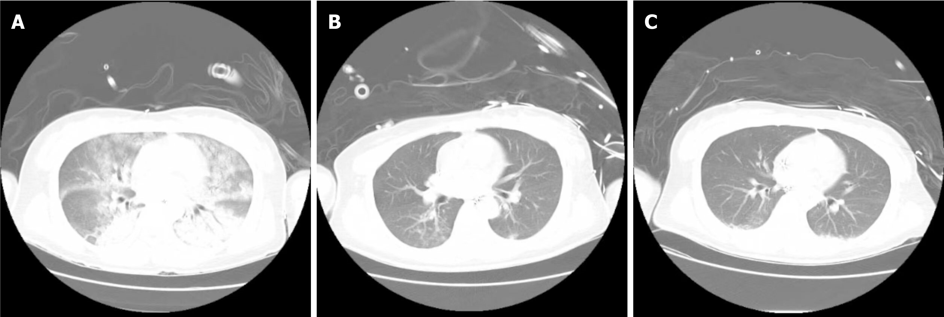
Figure 2 Chest computed tomography.
A: The first chest computed tomography (CT) examination considered pulmonary edema and lung infection; B: Preliminary consideration was that the patient had aspiration pneumonia. Chest CT was reexamined after 5 days of treatment with piperacillin sodium, and the inflammation was absorbed before; C: After extracorporeal membrane oxygenation and intra-aortic balloon counterpulsation withdrawal were withdrawn, a hot topic appeared, considering bac-teremia, adding vancomycin on the basis of using meropenem to resist infection, and re-examining chest CT inflammation before absorption.
- Citation: Zeng SY, Wu HH, Yu ZH, Zhang QQ. Extracorporeal membrane oxygenation combined with intra-aortic balloon counterpulsation for pheochromocytoma: A case report. World J Clin Cases 2025; 13(15): 102343
- URL: https://www.wjgnet.com/2307-8960/full/v13/i15/102343.htm
- DOI: https://dx.doi.org/10.12998/wjcc.v13.i15.102343


