Copyright
©The Author(s) 2025.
World J Clin Cases. May 26, 2025; 13(15): 101272
Published online May 26, 2025. doi: 10.12998/wjcc.v13.i15.101272
Published online May 26, 2025. doi: 10.12998/wjcc.v13.i15.101272
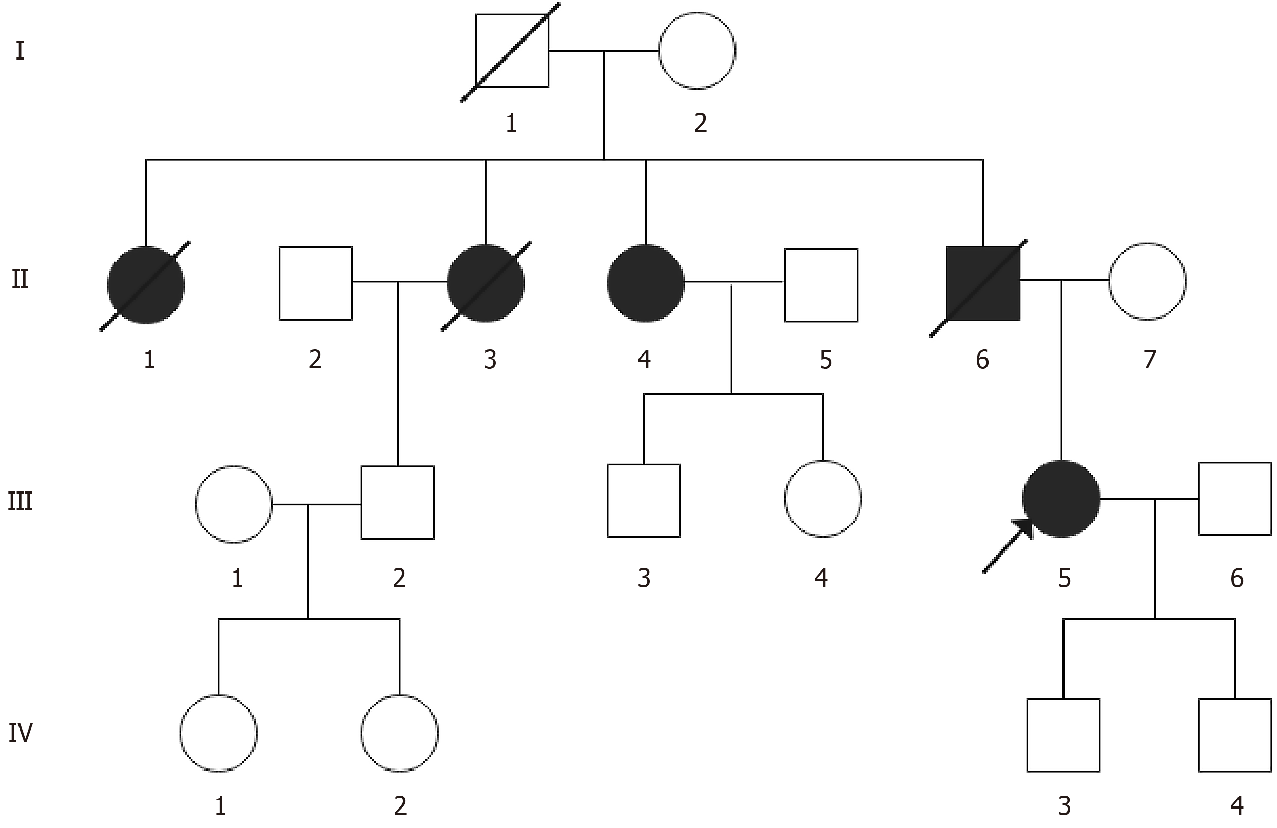
Figure 1 The pedigree chart of the family.
Box: Normal male; Circle: Normal female; Darkened: Affected; Slashed: Decreased; Arrow: The proband.
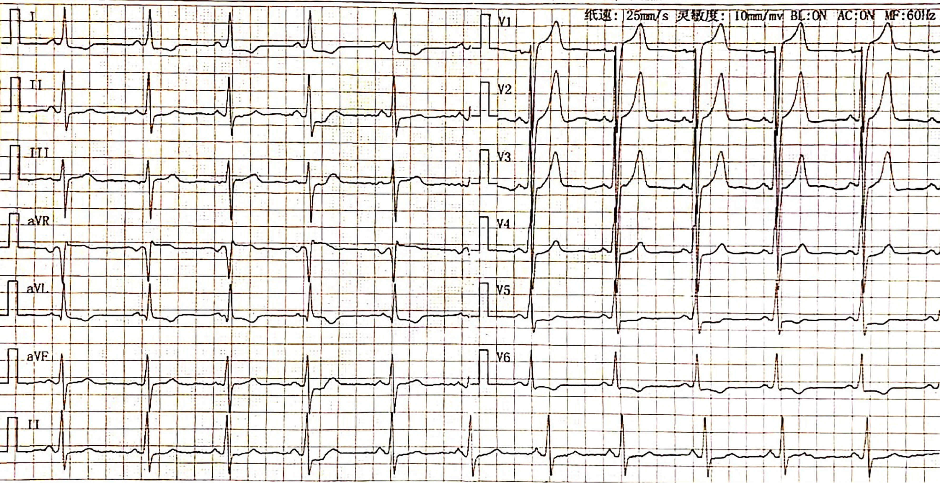
Figure 2
The electrocardiogram of the proband showed sinus rhythm and the S-T segment of leads I, avL and V6 was depressed and the T wave was inverted change.
Figure 3 The echocardiogram.
A: The first echocardiogram of the proband in 2018 was characterized by asymmetric thickening of left ventricle (LV) wall and thickening ventricular septum, without enlargement of LV (37 mm), on the horizontal long axis of LV, respectively; B: The second echocardiogram showed the enlargement of left atrium and LV (43 mm), the thinning of ventricular septum on the long axis of LV (The former is a short axis view of the papillary muscle, while the latter is a long axis view of the LV).
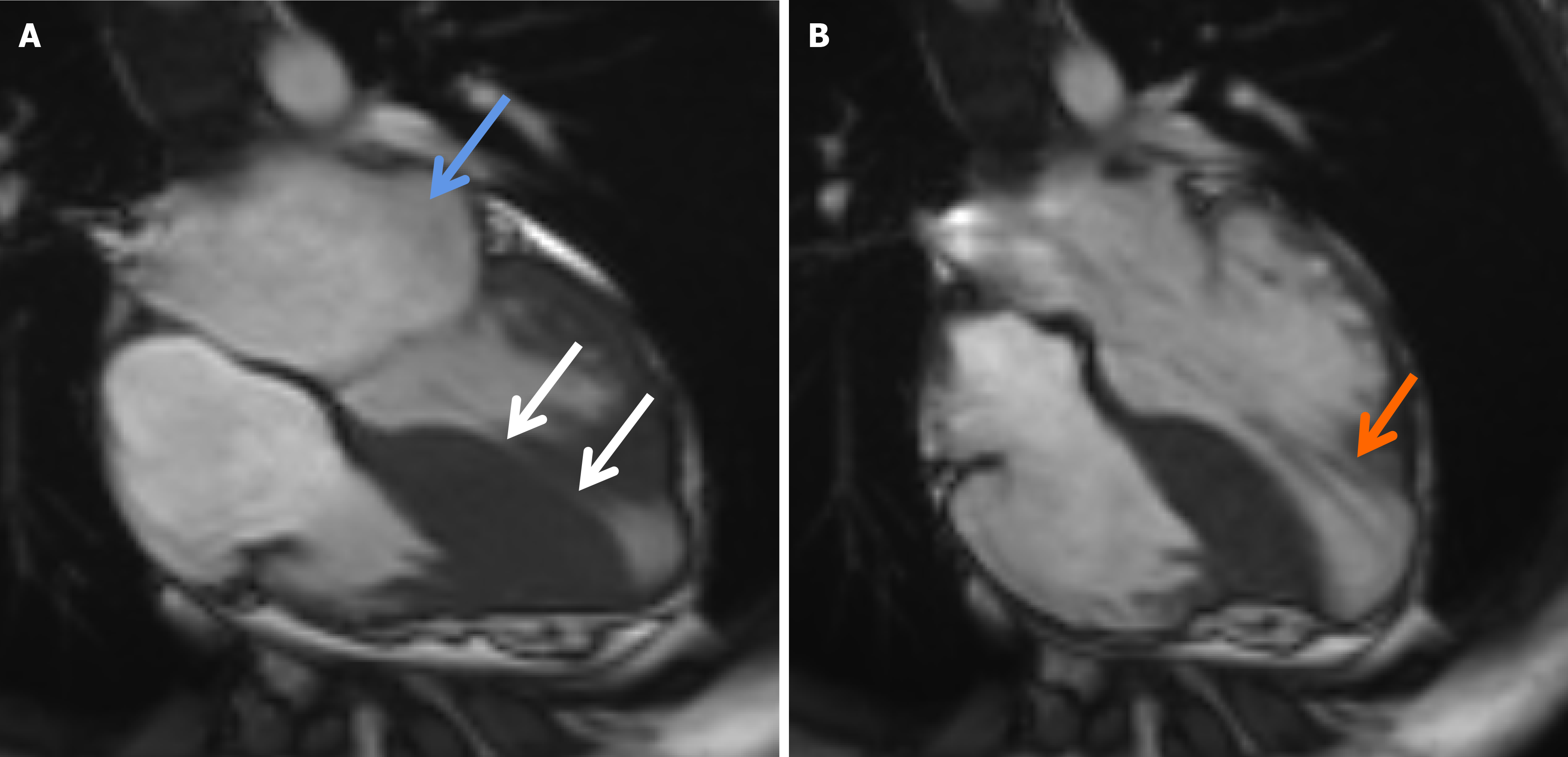
Figure 4 The cardiac magnetic resonance imaging findings of the proband.
A: The cardiac magnetic resonance imaging (CMR) found enlargement of left atrium (shown by the blue arrow), asymmetric hypertrophy of ventricular septum (shown by the white arrows), thinning of the bottom of ventricular septal base and left ventricle free wall; B: An apical ventricular aneurysm (about 24.6 mm × 29.8 mm) was shown by CMR (the orange arrow).
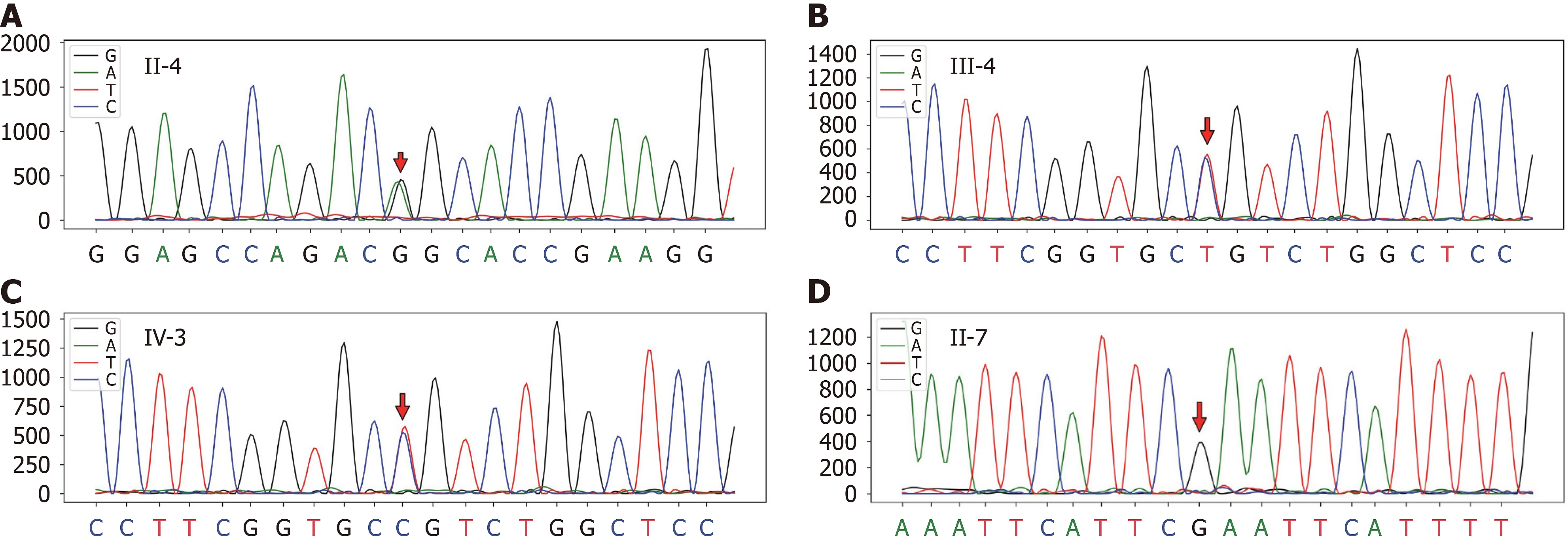
Figure 5 Sanger sequencing DNA chromatogram in MYH7 gene of the pedigree.
A: II-4; B: III-4; C: IV-3; D: II-7. Subjects (II-4, III-4 and IV-3) all presented with a heterozygous missense mutation (c.746G>A, p.R249Q). Subject II-7 was normal.
- Citation: Hong Y, Fan Z, Guo Y, Ma HH, Zeng SZ, Xi HT, Yang J, Luo K, Luo R, Li XP. MYH7 mutation in a pedigree with familial dilated hypertrophic cardiomyopathy: A case report and review of literature. World J Clin Cases 2025; 13(15): 101272
- URL: https://www.wjgnet.com/2307-8960/full/v13/i15/101272.htm
- DOI: https://dx.doi.org/10.12998/wjcc.v13.i15.101272


