Copyright
©The Author(s) 2025.
World J Clin Cases. May 16, 2025; 13(14): 103501
Published online May 16, 2025. doi: 10.12998/wjcc.v13.i14.103501
Published online May 16, 2025. doi: 10.12998/wjcc.v13.i14.103501
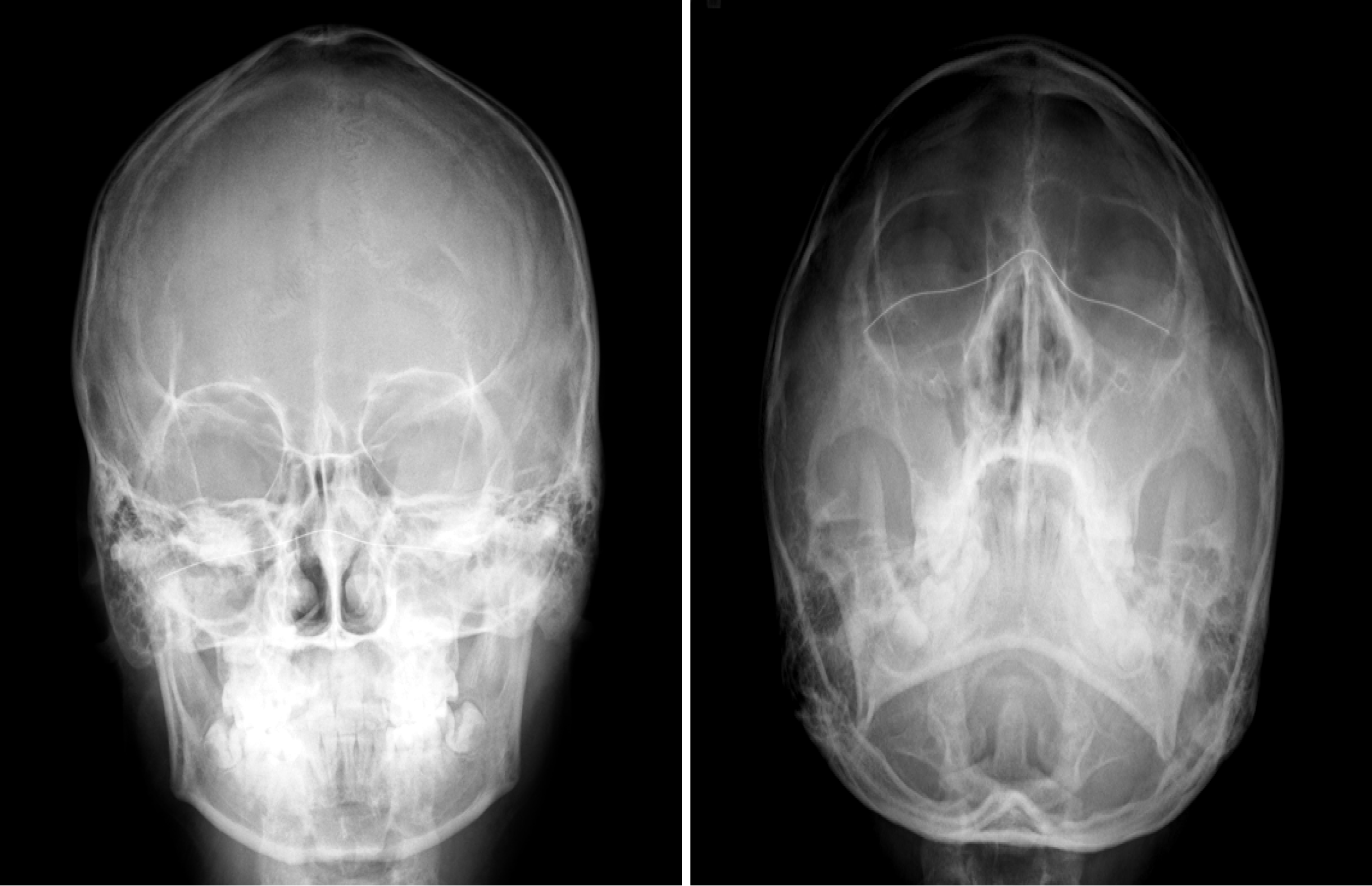
Figure 1
Film paranasal sinus showed mucoperiosteal thickening in the right maxillary sinus and total opacification in the left maxillary sinus.
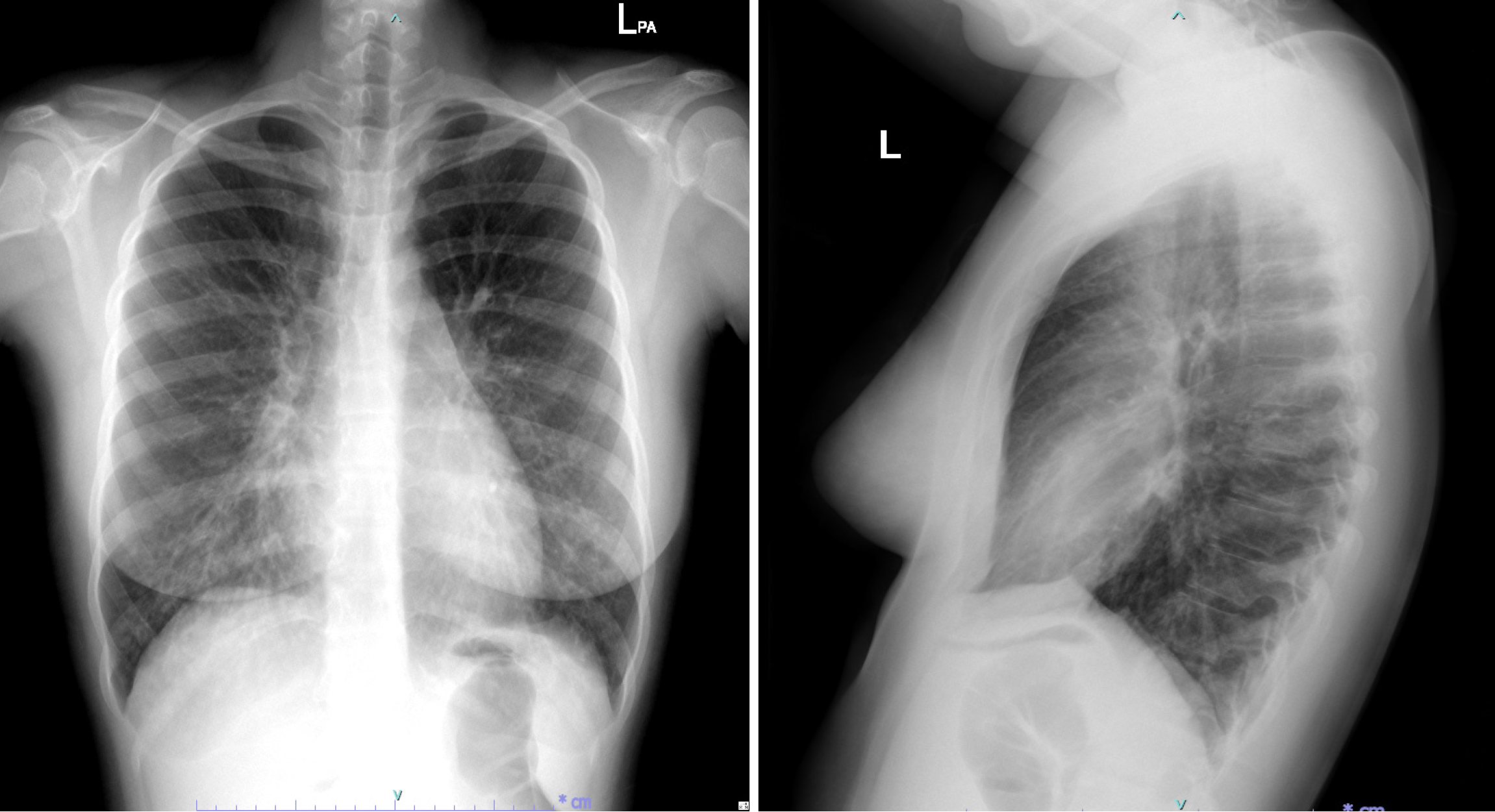
Figure 2
Chest radiograph showed reticulonodular infiltration in both middle to lower lung zones.
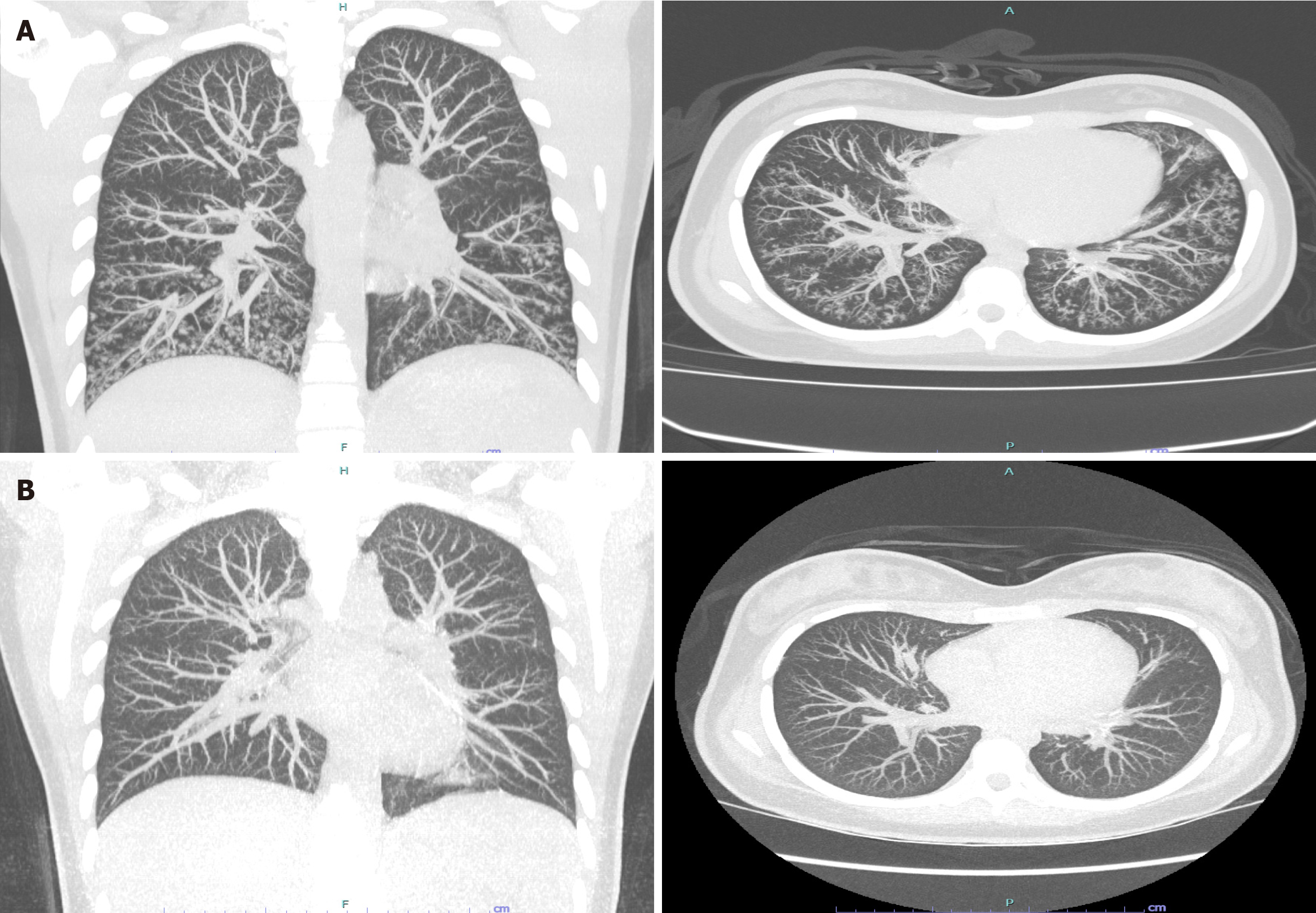
Figure 3 Chest high-resolution computed tomography under maximum intensity projection.
A: Before treatment, showed multiple centrilobular nodules with a tree-in-bud pattern; B: 10 months after treatment showed complete resolution of abnormal findings.
- Citation: Klubdaeng A, Tovichien P. Diffuse panbronchiolitis in children misdiagnosed as asthma: A case report. World J Clin Cases 2025; 13(14): 103501
- URL: https://www.wjgnet.com/2307-8960/full/v13/i14/103501.htm
- DOI: https://dx.doi.org/10.12998/wjcc.v13.i14.103501


