Copyright
©The Author(s) 2025.
World J Clin Cases. May 16, 2025; 13(14): 101950
Published online May 16, 2025. doi: 10.12998/wjcc.v13.i14.101950
Published online May 16, 2025. doi: 10.12998/wjcc.v13.i14.101950
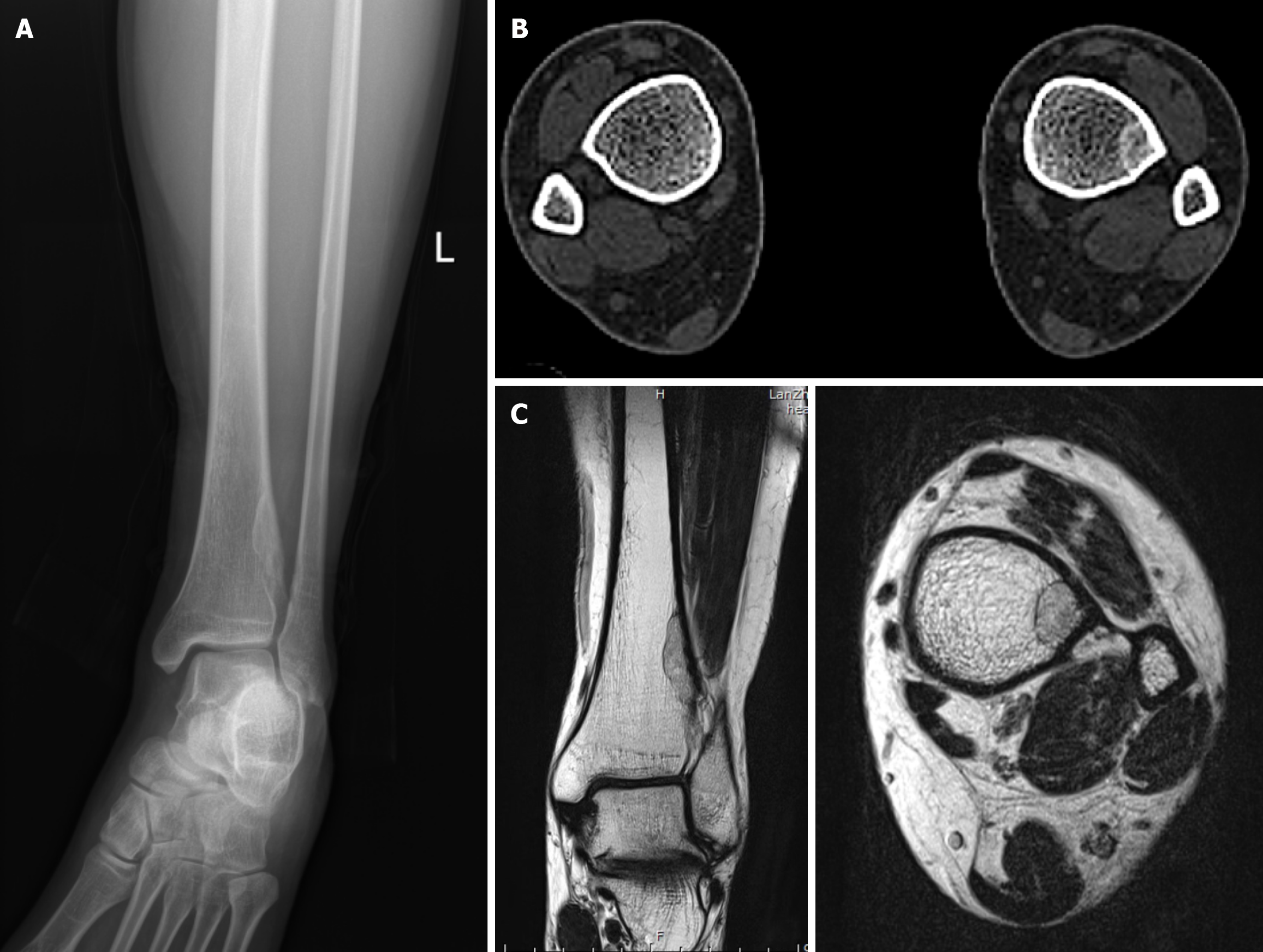
Figure 1 Imaging examination of the patient.
A: Preoperative X-ray showed that the lesion in the medial cortex of the left lower tibia showed an ellipsoidal increase in bone density, with a clear boundary and bone pattern; B: Preoperative computerized tomography showed an irregular ground-glass opacity in the left distal tibia, which measured about 24 mm × 8 mm in maximum diameter. There were slight thickening and sclerotic rim of the adjacent cortex; C: Preoperative magnetic resonance revealed an irregular slightly longer T1-weighted image and slightly longer T2-weighted image signal in the medial subcortical bone of the left distal tibial, with a uniform low signal ring at the edge of the left lower tibia, with a size of about 28 mm × 8 mm × 12 mm.
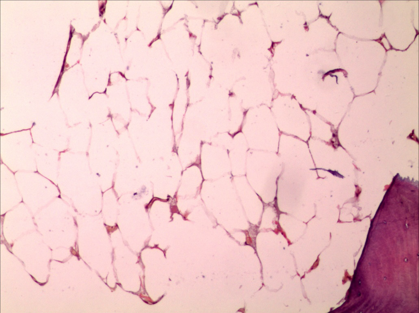
Figure 2
Postoperative pathological examination showed that there were a large number of mature fat cells in the bone cortex, with equal size and no obvious atypia, which were consistent with intraosseous lipoma (hematoxylin and eosin staining, × 100).
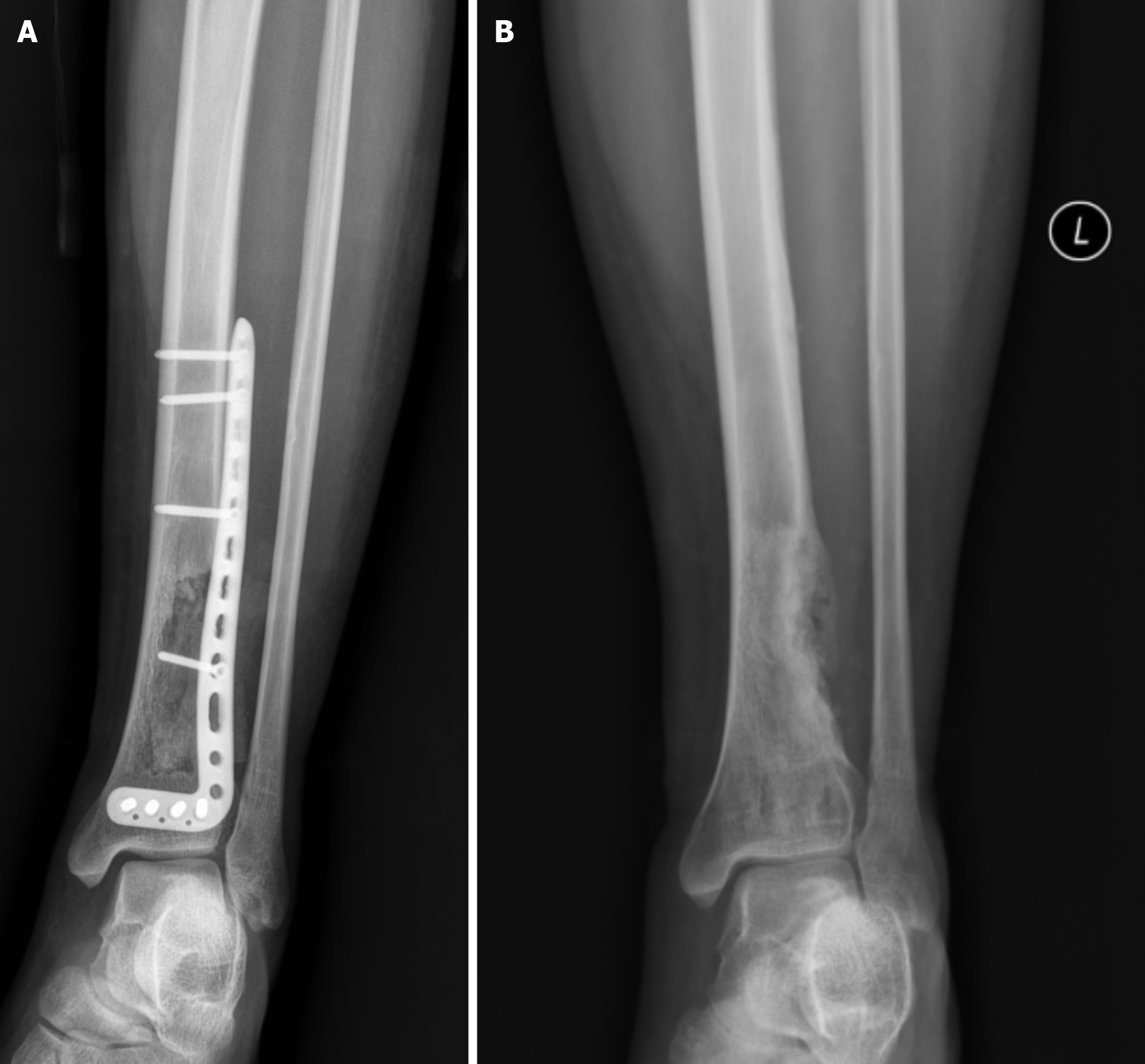
Figure 3 Postoperative X-ray of the patient.
A: Postoperative X-ray at 1 month showed that the lesion of the left lower tibia was curetted and internal fixation was in place; B: Postoperative X-ray at 30 months showed that multiple bone grafts were seen in the lesion defect area of the distal tibia on the left side.
- Citation: Liu P, Zhang K, Zeng JK, Chang YF, Zhuang KP, Zhou SH. Clinical, radiologic, and pathologic study of intraosseous lipoma of the tibia: A case report. World J Clin Cases 2025; 13(14): 101950
- URL: https://www.wjgnet.com/2307-8960/full/v13/i14/101950.htm
- DOI: https://dx.doi.org/10.12998/wjcc.v13.i14.101950


