Copyright
©The Author(s) 2025.
World J Clin Cases. May 6, 2025; 13(13): 102457
Published online May 6, 2025. doi: 10.12998/wjcc.v13.i13.102457
Published online May 6, 2025. doi: 10.12998/wjcc.v13.i13.102457
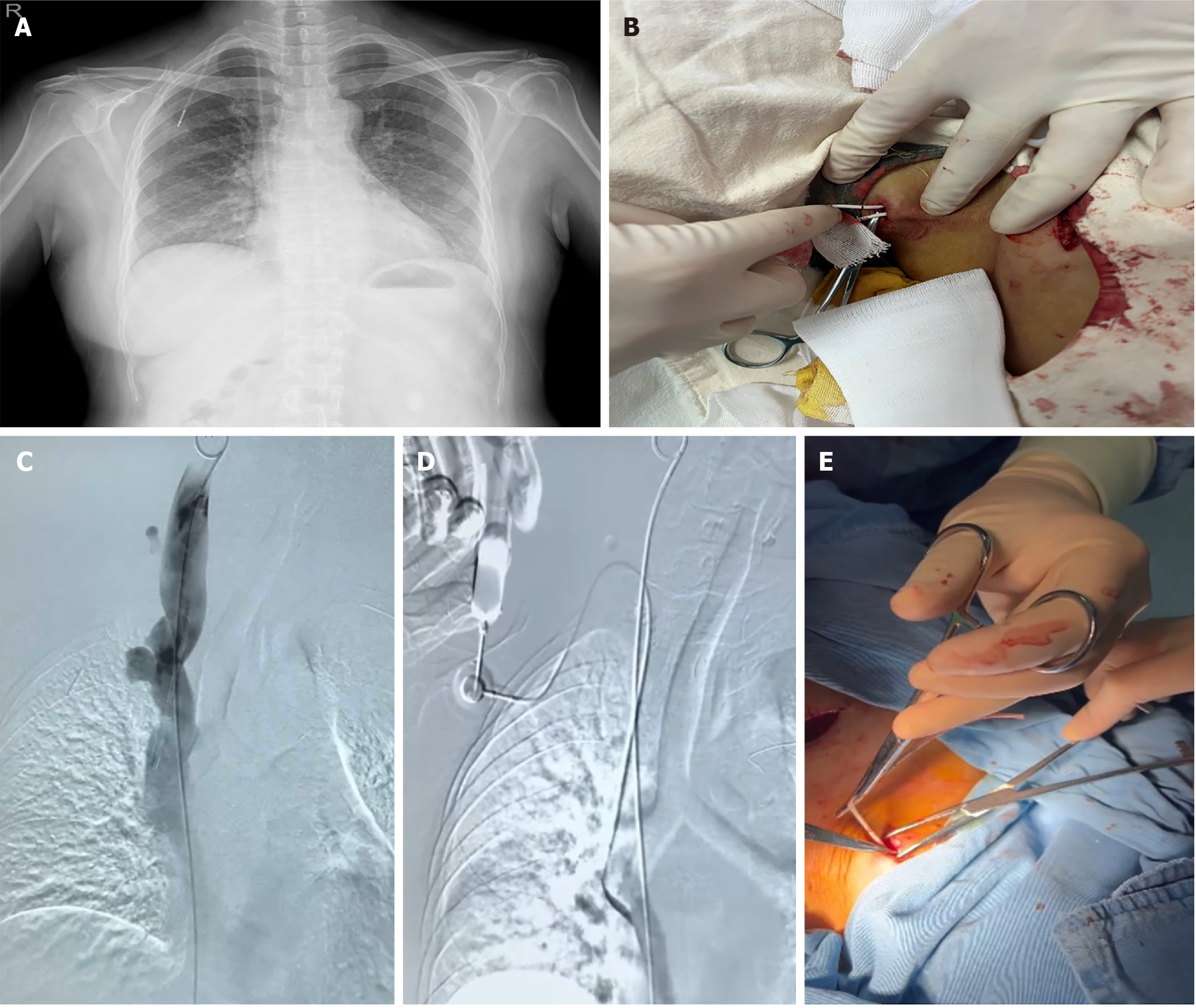
Figure 1 Investigation and surgical images related to the patient’s totally implantable venous access port.
A: Chest radiograph taken before totally implantable venous access port removal; B: Image showing the challenges encountered during catheter extraction through the neck incision; C: Digital subtraction angiography performed by inserting the catheter via femoral vein puncture into the internal jugular vein; D: Digital subtraction angiography image of contrast media injection from the totally implantable venous access port; E: Tissue separation around the catheter using a mosquito clamp.
- Citation: Chen J, Tang M, Han QY, Tang L, Yu TH, Zhao YP, He CW. Difficulty removing a totally implantable venous access port: A case report. World J Clin Cases 2025; 13(13): 102457
- URL: https://www.wjgnet.com/2307-8960/full/v13/i13/102457.htm
- DOI: https://dx.doi.org/10.12998/wjcc.v13.i13.102457


