Copyright
©The Author(s) 2025.
World J Clin Cases. May 6, 2025; 13(13): 102108
Published online May 6, 2025. doi: 10.12998/wjcc.v13.i13.102108
Published online May 6, 2025. doi: 10.12998/wjcc.v13.i13.102108
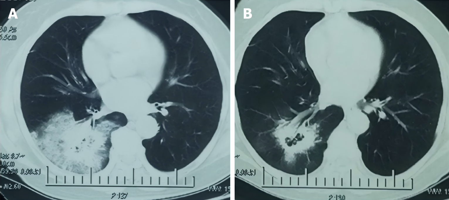
Figure 1 Chest computed tomography images reveal a mass in the lower lobe of the right lung.
A: Lung window at initial discovery; B: After anti-infective treatment.
Figure 2 Hem-o-lock clip.
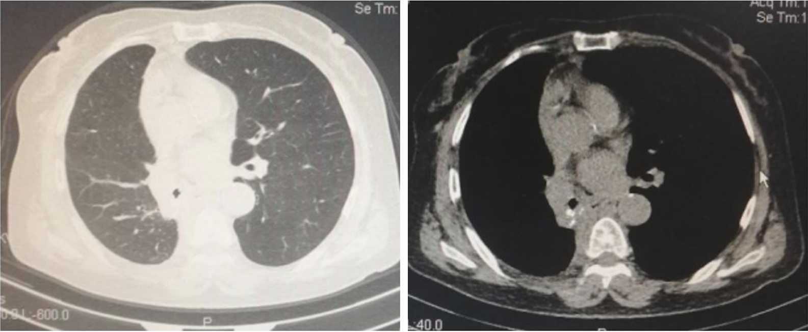
A and B: Lung window and mediastinum window after placement of the pigtail catheter under ultrasound guidance, respectively. The arrow in B indicates the high-density shadow which is the pigtail catheter; C: Hemolock clip expectorated orally.
Figure 3 One month after insertion of the chest tube, chest computed tomography imaging revealed good lung expansion without evidence of cavity or effusion, suggesting stabilization of the bronchial stump.
- Citation: Li QY, Wang XL, Zhang F, Wei HT. Bronchopleural fistula following application of Hem-o-lock clip at bronchial stump after lobectomy: A case report. World J Clin Cases 2025; 13(13): 102108
- URL: https://www.wjgnet.com/2307-8960/full/v13/i13/102108.htm
- DOI: https://dx.doi.org/10.12998/wjcc.v13.i13.102108


