Copyright
©The Author(s) 2025.
World J Clin Cases. May 6, 2025; 13(13): 101444
Published online May 6, 2025. doi: 10.12998/wjcc.v13.i13.101444
Published online May 6, 2025. doi: 10.12998/wjcc.v13.i13.101444
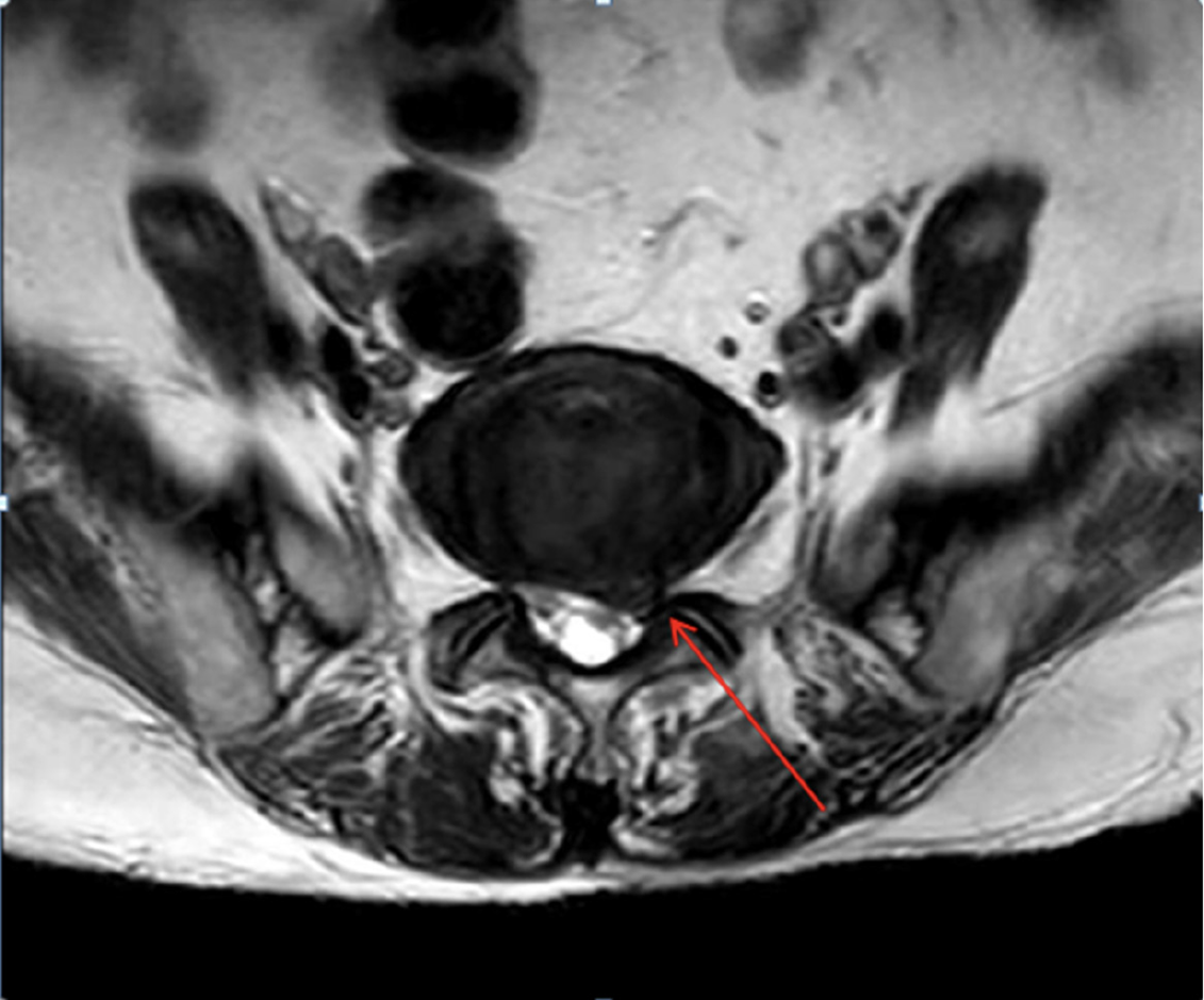
Figure 1 Axial section of a magnetic resonance imaging of the lumbar spine in L5/S1 showing a left paramedian disc herniation.
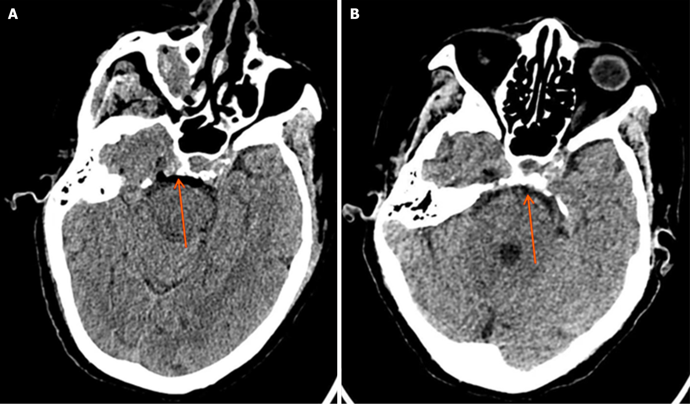
Figure 2 Computed tomography findings.
A: Axial section of a cerebral computed tomography (CT) scan on the operative day showing the pneumocephalus surrounding the brainstem; B: Axial section of a cerebral CT scan the following day showing modification of the pneumocephalus.
- Citation: Han C, Ren ZY, Jiang ZH, Luo YF. Cerebral complications after unilateral biportal endoscopic surgery: A case report. World J Clin Cases 2025; 13(13): 101444
- URL: https://www.wjgnet.com/2307-8960/full/v13/i13/101444.htm
- DOI: https://dx.doi.org/10.12998/wjcc.v13.i13.101444


