Copyright
©The Author(s) 2025.
World J Clin Cases. Apr 16, 2025; 13(11): 98979
Published online Apr 16, 2025. doi: 10.12998/wjcc.v13.i11.98979
Published online Apr 16, 2025. doi: 10.12998/wjcc.v13.i11.98979
Figure 1 Computed tomography examination of the patient.
A: On August 23, 2023, enhanced chest computed tomography (CT) revealed a soft-tissue shadow that was visible on the right side of the anterior mediastinum, with a size of approximately 78 mm × 53 mm. After enhancement, the mass was uneven and mildly enhanced, the surrounding fat space was blurred, and the superior vena cava and the right atrium were compressed; B: On August 28, 2023, chest CT revealed a soft-tissue shadow that was visible on the right side of the anterior mediastinum; the size of the mass was approximately 64 mm × 55 mm, the surrounding fat space was blurred, and the superior vena cava and the right atrium were compressed; C: On September 11, 2023, chest CT revealed a soft-tissue shadow that was visible on the right side of the anterior mediastinum; the size of the mass was approximately 39 mm × 54 mm, the surrounding fat space was blurred, and the sizes and shapes of the large blood vessels of the heart were normal.
Figure 2 Preoperative pathology.
Preoperative pathology revealed a small amount of lymphoid tissue and epithelioid cells with necrosis. When combined with immunohistochemical results, which were consistent with thymoma, and considering that there was little tissue, it was recommended that specimens be further classified after surgery. The immunohistochemistry results were as follows: TdT (partial +), CD5 (partial +), CD20 (small +), CD3 (partial +), CK (AE1/AE3) (+), CK19 (+), p63 (+), Ki-67 (20% +), CDla (partial +), CD117 (individual +), CK (+), and p53 (minor +). HE: Hematoxylin and eosin.
Figure 3 Postoperative pathology.
Postoperative pathology revealed anterior mediastinal thymoma (type B2), cyst formation in some areas, fibrosis and large necrosis, and infiltration of the capsule. Immunohistochemistry revealed the following: CK (AE1/AE3) (+), CK19 (+), CK5/6 (+), p63 (+), Ki-67 (+), TdT (-), CD5 (-), CD20 (-), CD3 (-), CDla (-), and CD117 (-). HE: Hematoxylin and eosin.
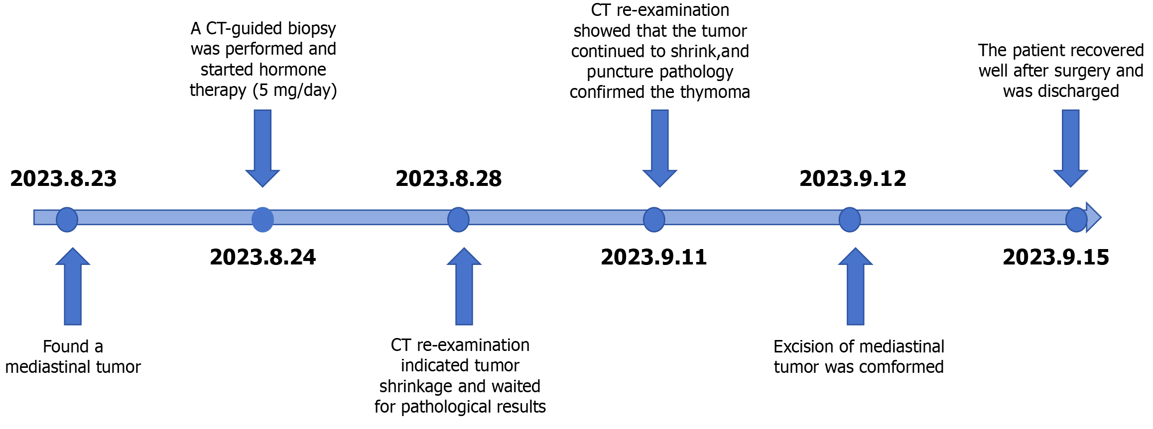
Figure 4 Patient treatment flowchart.
CT: Computed tomography.
- Citation: Yao JK, He ZY, Zhu Z, Huang HT. Treatment of thymoma with low-dose glucocorticoids before surgery for significant tumor shrinkage: A case report. World J Clin Cases 2025; 13(11): 98979
- URL: https://www.wjgnet.com/2307-8960/full/v13/i11/98979.htm
- DOI: https://dx.doi.org/10.12998/wjcc.v13.i11.98979


