Copyright
©The Author(s) 2025.
World J Clin Cases. Apr 16, 2025; 13(11): 98570
Published online Apr 16, 2025. doi: 10.12998/wjcc.v13.i11.98570
Published online Apr 16, 2025. doi: 10.12998/wjcc.v13.i11.98570
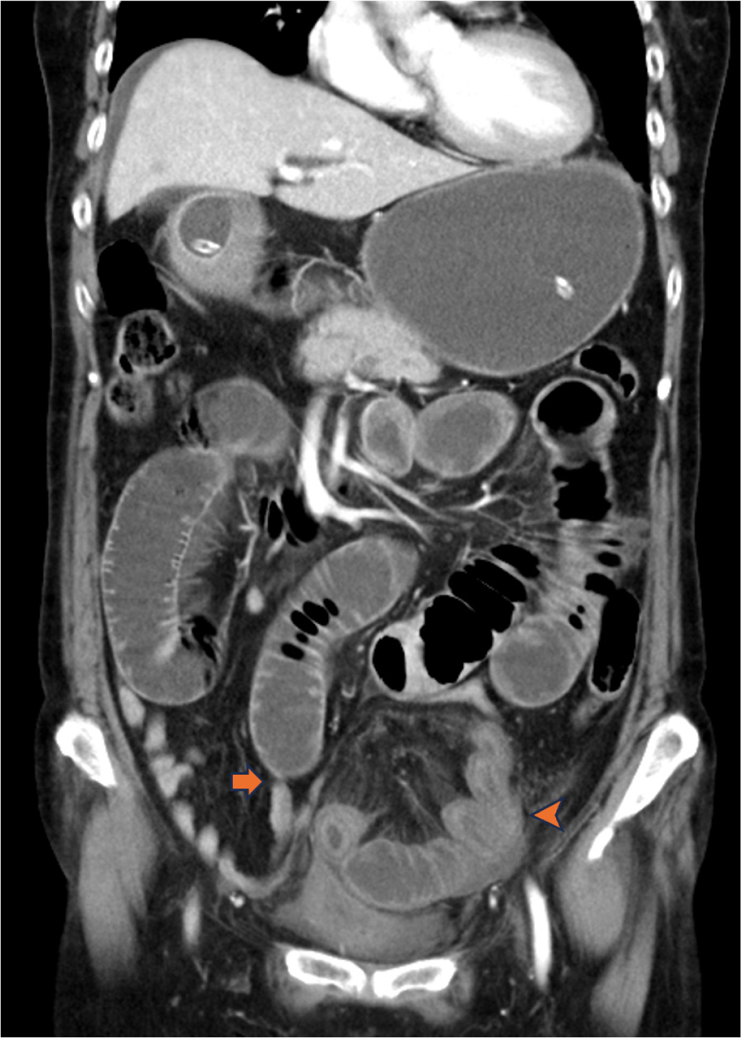
Figure 1 Pre-operative abdominal computed tomography scan.
Abdominal computed tomography revealed segmental dilatation of the small bowel with a transitional zone (arrow), as well as a segment of suspicious closed loop of the small bowel (arrowhead).
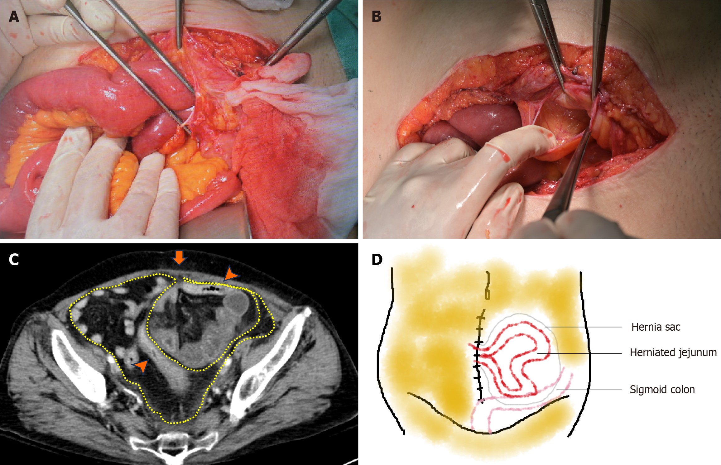
Figure 2 Findings of parietal peritoneal hernia.
A: After laparotomy, a large segment of distal jejunum was found herniated into the retrorectus preperitoneal space; B: After reduction of the herniated intestine, a complete hernia sac was observed through a peritoneal defect on the prior scar; C: Pre-operative computed tomography showed the hernia sac located between the intact rectus abdominis (arrow) and sigmoid colon (arrowhead, representing part of the dome of the sac). The yellow line indicates the parietal peritoneum; D: This perspective drawing illustrates the spatial relationship of the hernia in a simple way.
- Citation: Chou YC. Parietal peritoneal hernia after abdominal hysterectomy for forty years: A case report. World J Clin Cases 2025; 13(11): 98570
- URL: https://www.wjgnet.com/2307-8960/full/v13/i11/98570.htm
- DOI: https://dx.doi.org/10.12998/wjcc.v13.i11.98570


