Copyright
©The Author(s) 2025.
World J Clin Cases. Apr 16, 2025; 13(11): 101668
Published online Apr 16, 2025. doi: 10.12998/wjcc.v13.i11.101668
Published online Apr 16, 2025. doi: 10.12998/wjcc.v13.i11.101668
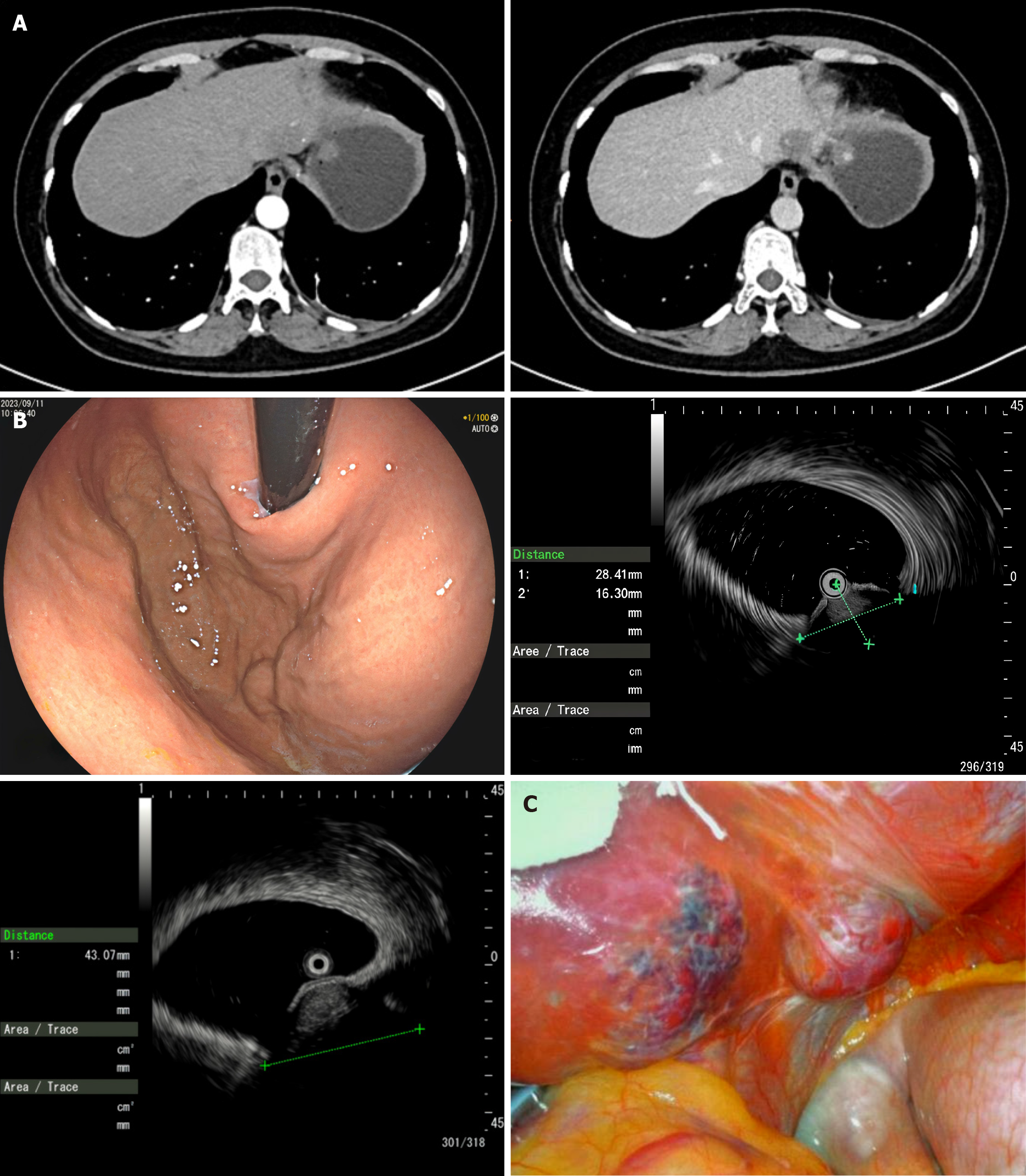
Figure 1 The patient’s imaging examination and laparoscopy.
A: Enhanced computed tomography image of two circular nodules in the fundus of the stomach; B: Endoscopic ultrasound image of two connected spherical bulges in the anterior wall of the fundus of the stomach; C: Laparoscopic visualization of two unexpected lesions in the left lobe of the liver.
- Citation: Wang JZ, Chen H. Hepatic hemangiomas mimicking gastrointestinal stromal tumors: A case report. World J Clin Cases 2025; 13(11): 101668
- URL: https://www.wjgnet.com/2307-8960/full/v13/i11/101668.htm
- DOI: https://dx.doi.org/10.12998/wjcc.v13.i11.101668


