Copyright
©The Author(s) 2025.
World J Clin Cases. Apr 6, 2025; 13(10): 97584
Published online Apr 6, 2025. doi: 10.12998/wjcc.v13.i10.97584
Published online Apr 6, 2025. doi: 10.12998/wjcc.v13.i10.97584
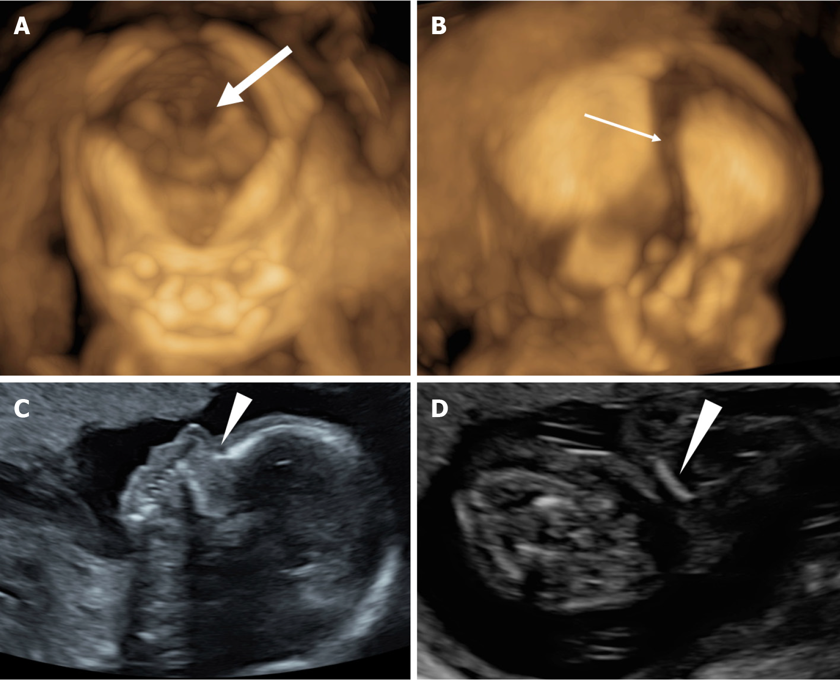
Figure 1 Two-dimensional and three-dimensional ultrasound of fetal cleidocranial dysplasia.
A: Three-dimensional (3D) ultrasonography showed enlargement of the anterior fontanelle (thick arrow); B: 3D ultrasound showed temporal suture widening (thin arrow); C: 2D ultrasound showed the absence of nasal bone (short arrowhead); D: 2D ultrasound showed that the clavicle was short and straight without a typical “S” shape (long arrowhead).
Figure 2 Sequencing results show that the fetus carried pathogenic mutations in the RUNX2 gene (c.
674G>A) (black arrow).
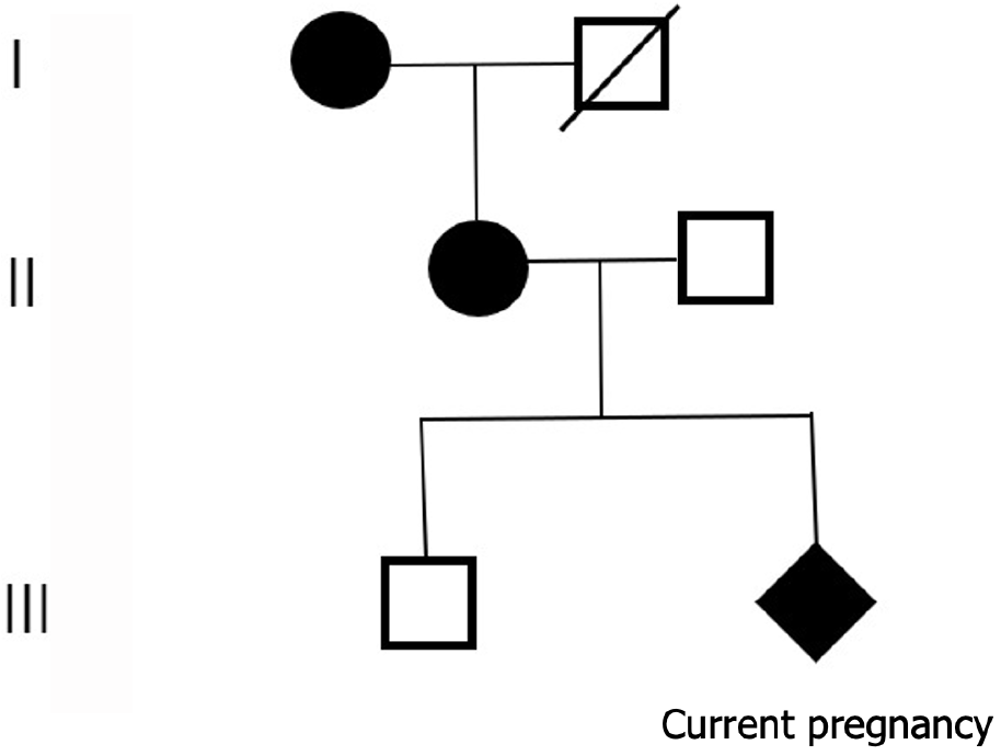
Figure 3 Pedigree of the affected family.
Circles and squares represent females and males, respectively. Affected individuals are shown in black. Diamond represents current pregnancy. Slash represents death.
- Citation: Wang F, Dai PF, Gao WJ. Prenatal ultrasonography and genetic analysis of fetal cleidocranial dysplasia: A case report. World J Clin Cases 2025; 13(10): 97584
- URL: https://www.wjgnet.com/2307-8960/full/v13/i10/97584.htm
- DOI: https://dx.doi.org/10.12998/wjcc.v13.i10.97584


