Copyright
©The Author(s) 2025.
World J Clin Cases. Apr 6, 2025; 13(10): 101796
Published online Apr 6, 2025. doi: 10.12998/wjcc.v13.i10.101796
Published online Apr 6, 2025. doi: 10.12998/wjcc.v13.i10.101796
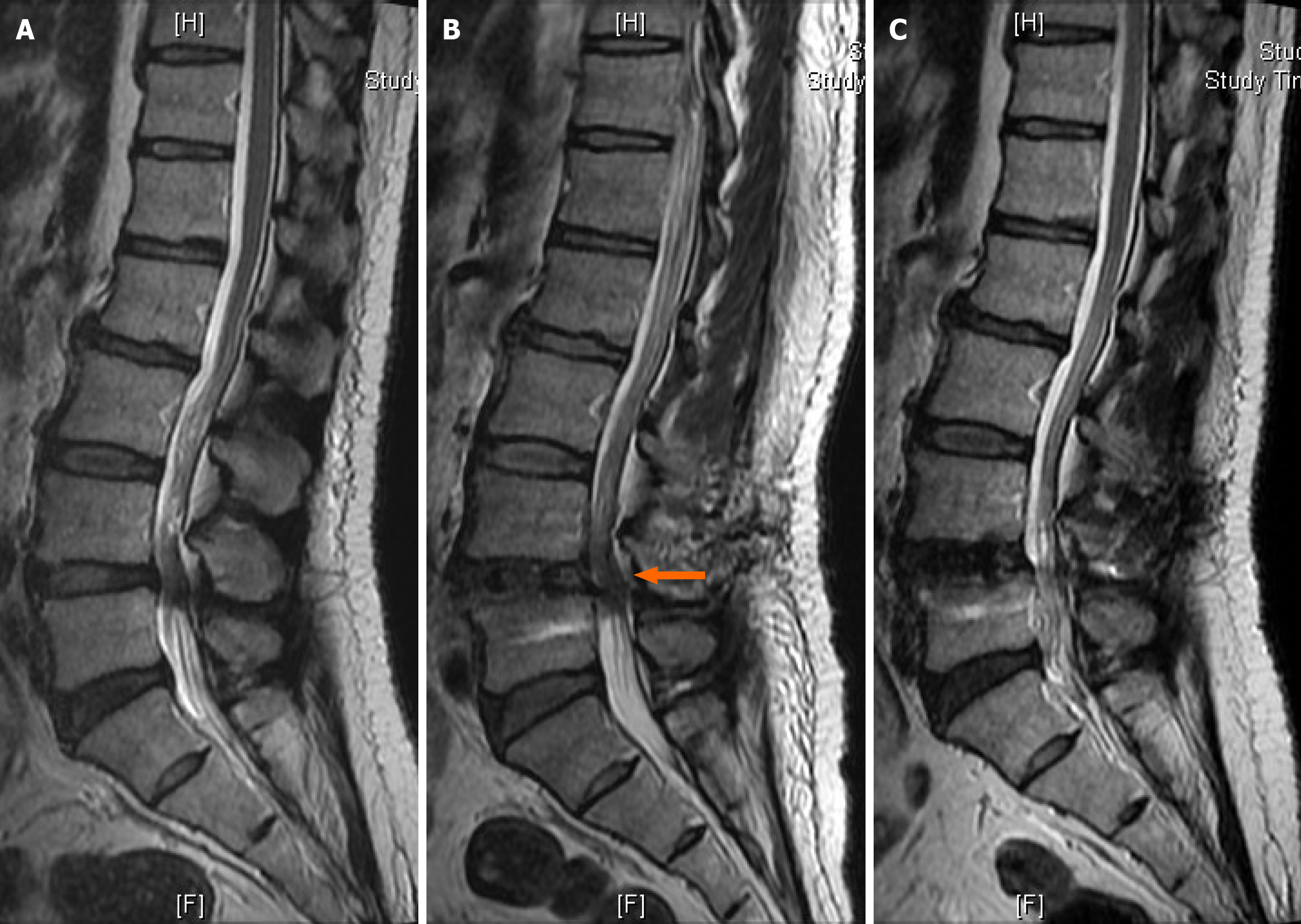
Figure 1 Magnetic resonance imaging findings before and after spinal surgery.
A: Preoperative magnetic resonance imaging (MRI); B: Postoperative day5 MRI showed decreased diameter of the spinal canal at L4-5 level with spinal stenosis, suspected local hematoma (arrow); C: MRI at 3 months later after the repeated exploration.
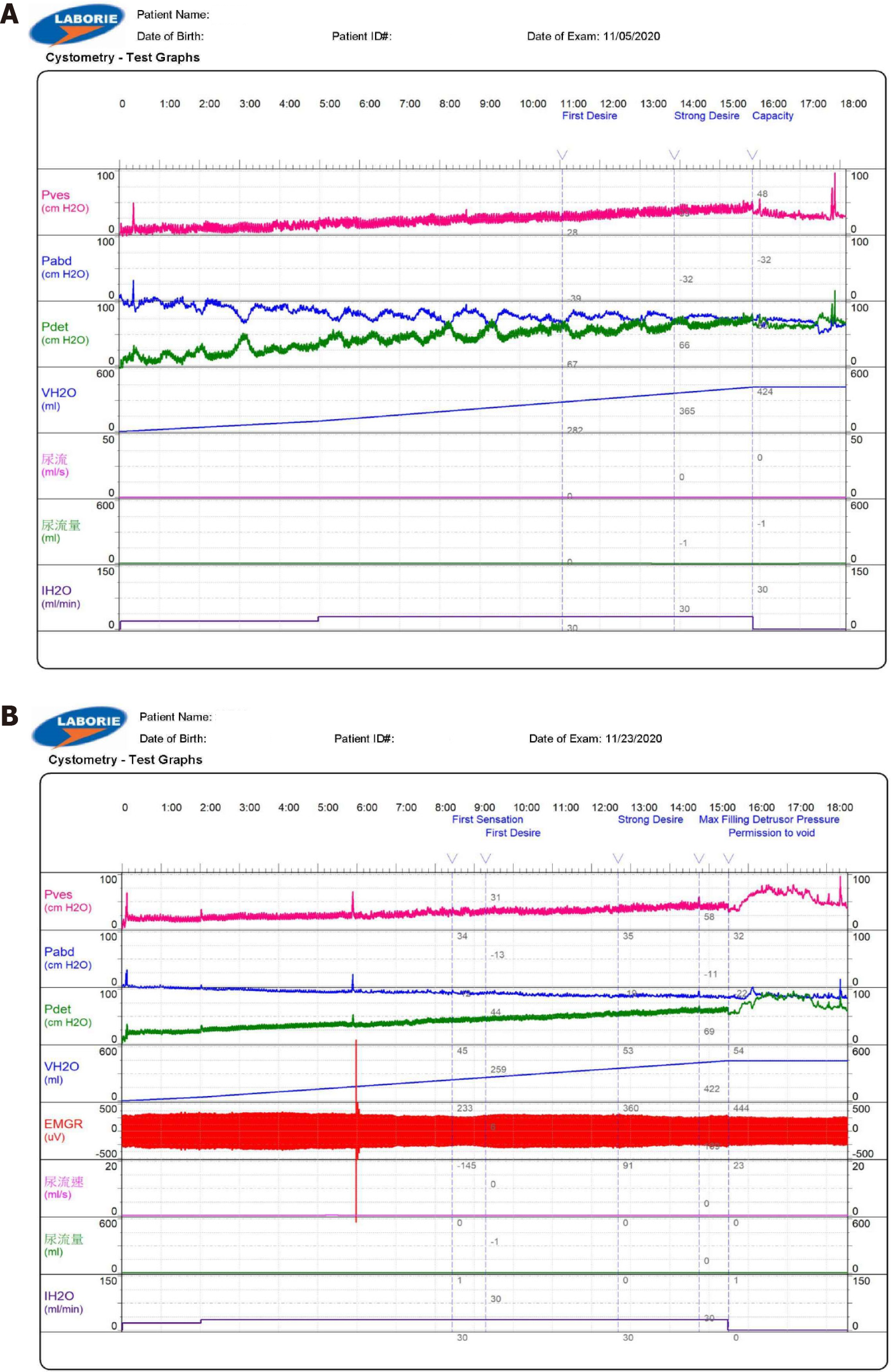
Figure 2 Postoperative cystometrogram.
A: Postoperative cystometrogram at day 7 showed no bladder contraction even until the capacity to 424 mL and delay first sensation; B: Postoperative cystometrogram at 3 weeks showed some bladder contraction but high compliance.
- Citation: Yang KW, Lai WH, Huang DW. Cauda equina syndrome with urinary retention as a postoperative complication of lumbar spine surgery: A case report. World J Clin Cases 2025; 13(10): 101796
- URL: https://www.wjgnet.com/2307-8960/full/v13/i10/101796.htm
- DOI: https://dx.doi.org/10.12998/wjcc.v13.i10.101796


