Copyright
©The Author(s) 2025.
World J Clin Cases. Jan 6, 2025; 13(1): 99938
Published online Jan 6, 2025. doi: 10.12998/wjcc.v13.i1.99938
Published online Jan 6, 2025. doi: 10.12998/wjcc.v13.i1.99938
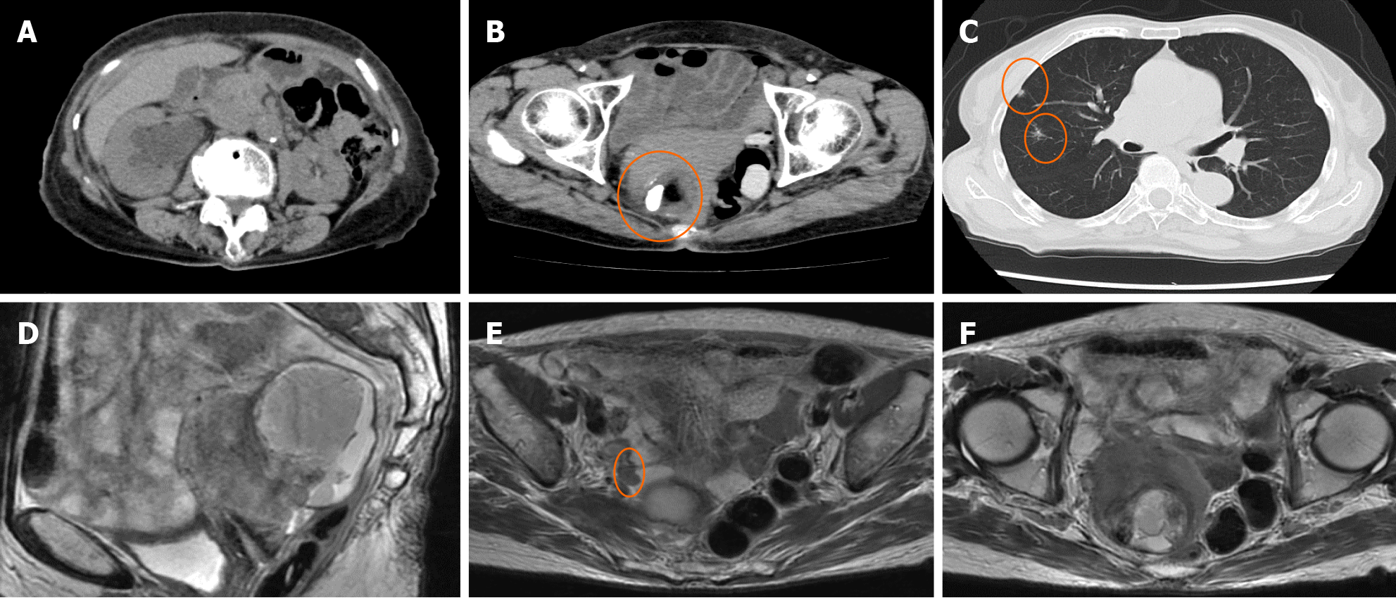
Figure 1 Computed tomography and magnetic resonance imaging findings.
A-C: Computed tomography revealed right hydronephrosis, a 6-cm solid cystic mass with fat and calcification densities, and multiple nodules in the lungs (orange circles); D-F: Magnetic resonance imaging showed that the tumor was adhering to the uterus and rectum. There was suspected dissemination near the right ureter (orange circle).
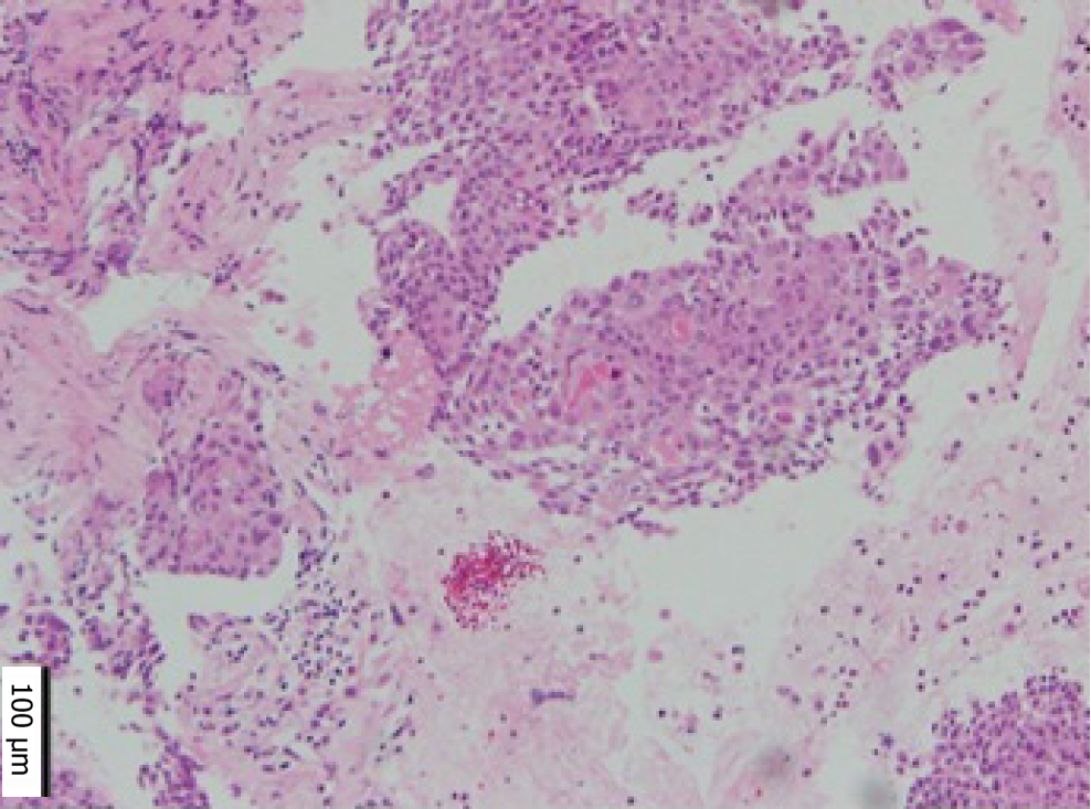
Figure 2 Pathological diagnosis using endoscopic ultrasound-guided fine-needle biopsy was squamous cell carcinoma.
Figure 3 Timeline of this case.
CCRT: Concurrent chemoradiotherapy.
- Citation: Kondo S, Suzuki T, Yoshiike K, Yamanaka S, Sonehara K, Nabeshima H, Oguchi O. Stage IV malignant transformation of mature cystic teratoma palliatively treated with concurrent chemoradiotherapy: A case report. World J Clin Cases 2025; 13(1): 99938
- URL: https://www.wjgnet.com/2307-8960/full/v13/i1/99938.htm
- DOI: https://dx.doi.org/10.12998/wjcc.v13.i1.99938


