Copyright
©The Author(s) 2024.
World J Clin Cases. Mar 26, 2024; 12(9): 1677-1684
Published online Mar 26, 2024. doi: 10.12998/wjcc.v12.i9.1677
Published online Mar 26, 2024. doi: 10.12998/wjcc.v12.i9.1677
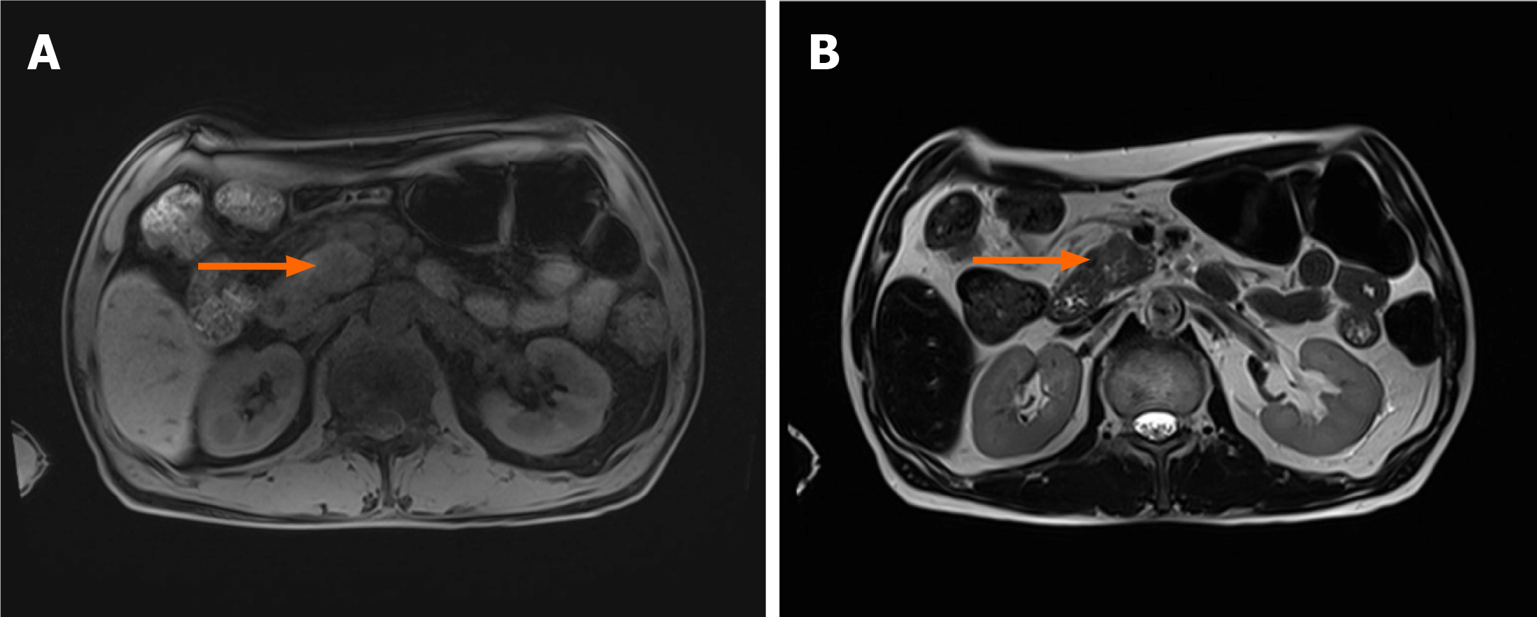
Figure 1 Magnetic resonance imaging.
A and B: An approximately 3 cm, ill-defined, space-occupying lesion in the inferior aspect of the head with high signal intensity on T1- and T2-weighted imaging (orange arrows).
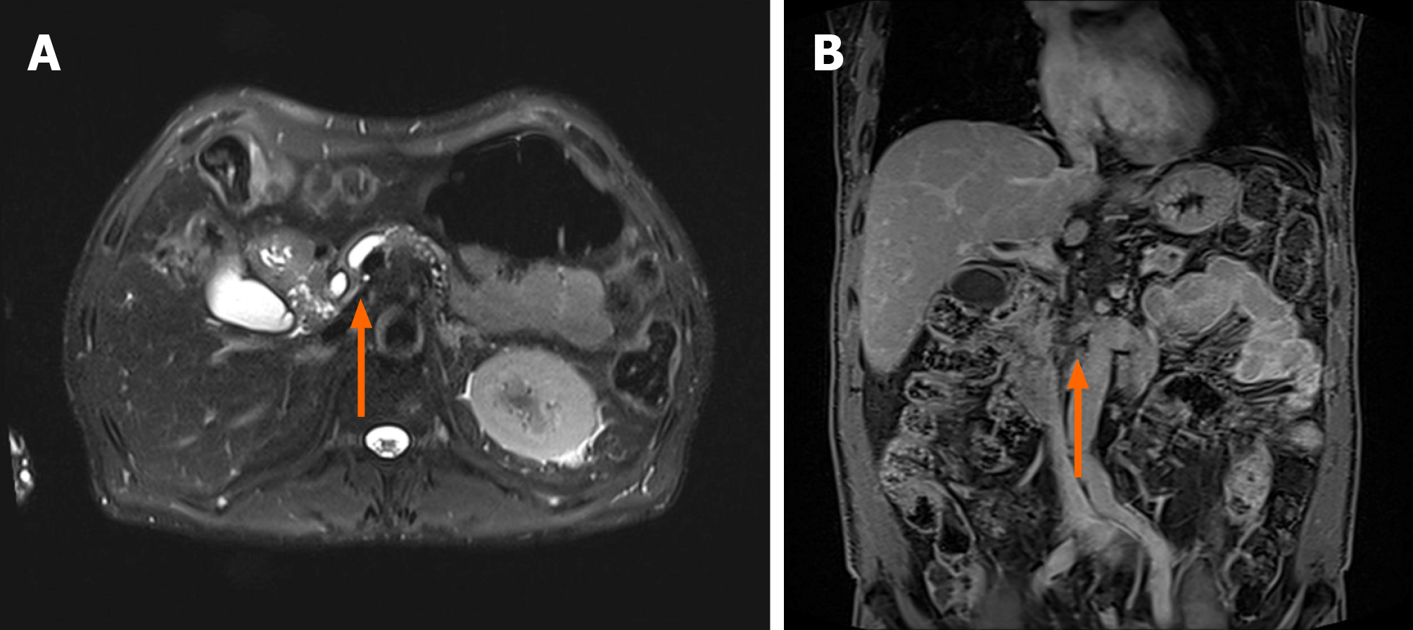
Figure 2 Magnetic resonance imaging.
A: An ill-defined space-occupying lesion invading the distal common bile duct and main pancreatic duct, both of which were narrowed, and the upstream main pancreatic duct dilated (orange arrow); B: The lesion severely encased the superior mesenteric artery (orange arrow).
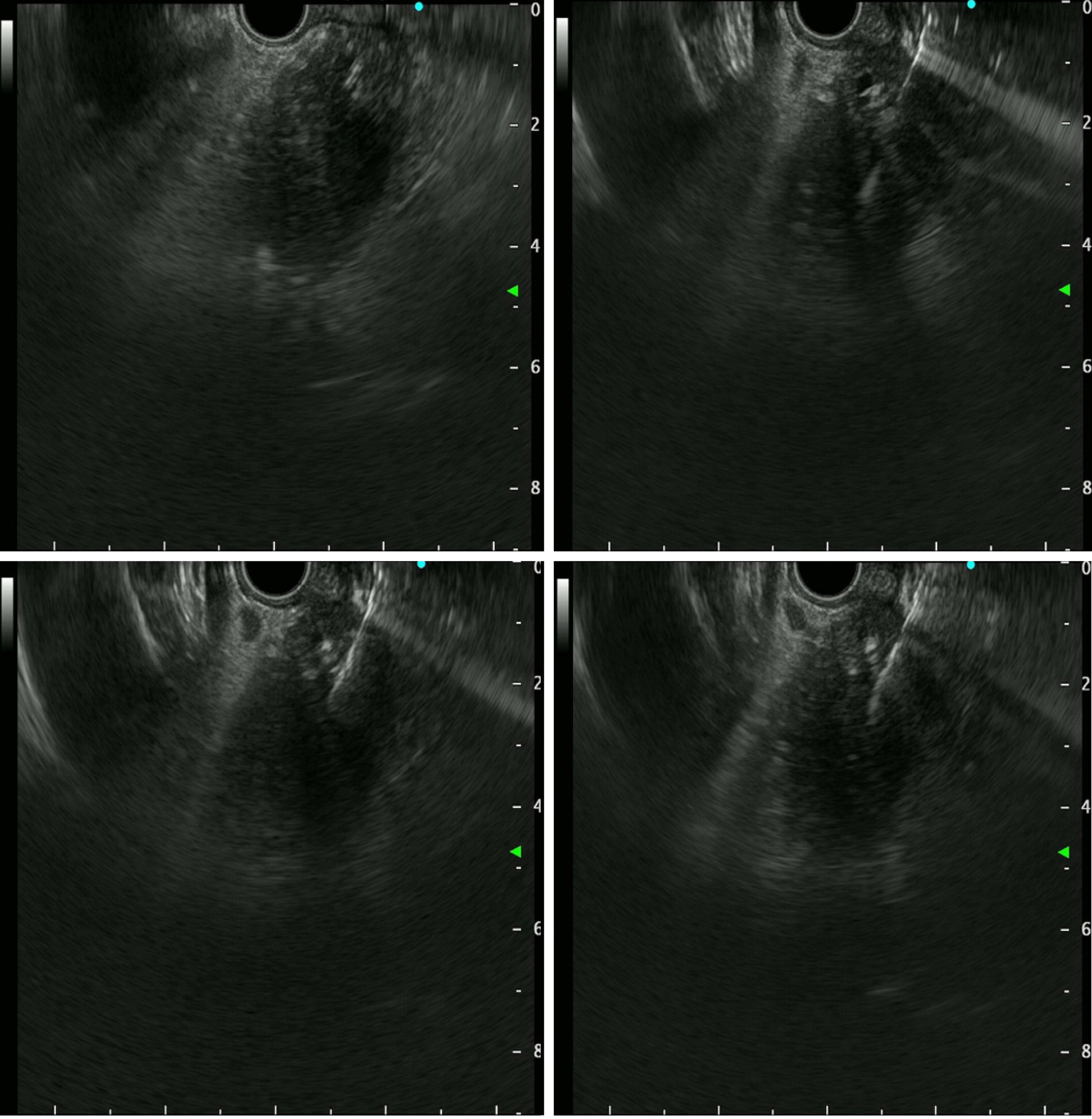
Figure 3 Endoscopic ultrasound-guided tissue sampling.
Endoscopic ultrasound (EUS) showed a 36.0 mm × 32.9 mm sized ill-defined relative heterogeneous hypoechoic mass in the pancreatic head. EUS-guided tissue sampling (EUS-TS) was performed using a 22-gauge EZ Shot (EZ Shot 3 Plus; Olympus, Tokyo, Japan) using the fanning method.
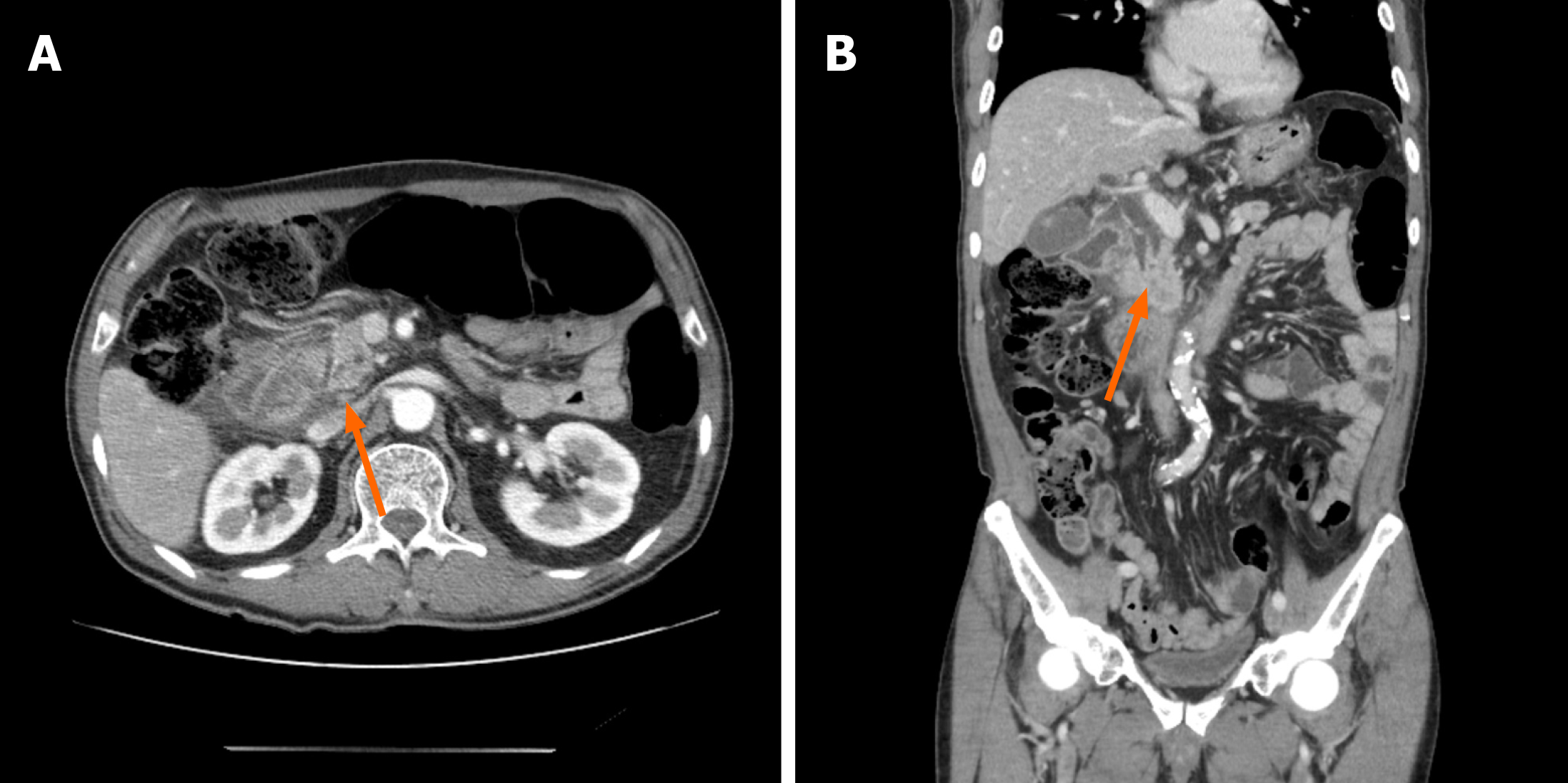
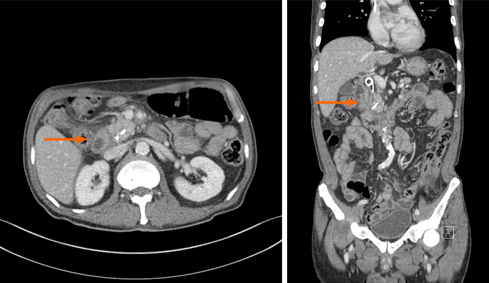
Figure 4 Computed tomography of the abdomen two days after endoscopic ultrasound-guided tissue sampling.
A and B: Edematous wall thickening of the second portion of the duodenum with adjacent fluid collection was detected and a leak was suspected (orange arrows).
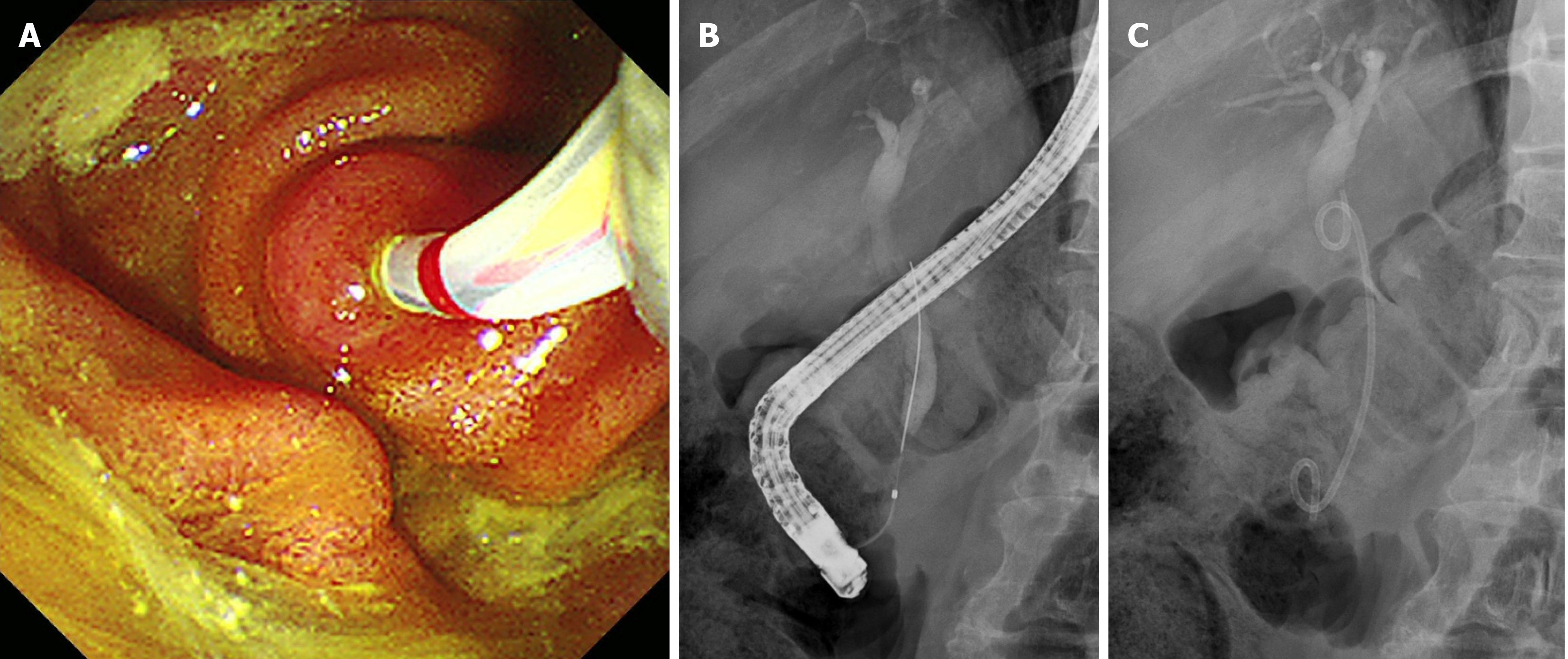
Figure 5 First endoscopic retrograde cholangiopancreatography.
A: Selective biliary cannulation was achieved only through the ampulla of Vater; B: There is no definitive dye leakage from the distal common bile duct; C: A 7F 7 cm-sized double pigtail plastic stent was deployed for effective biliary drainage.
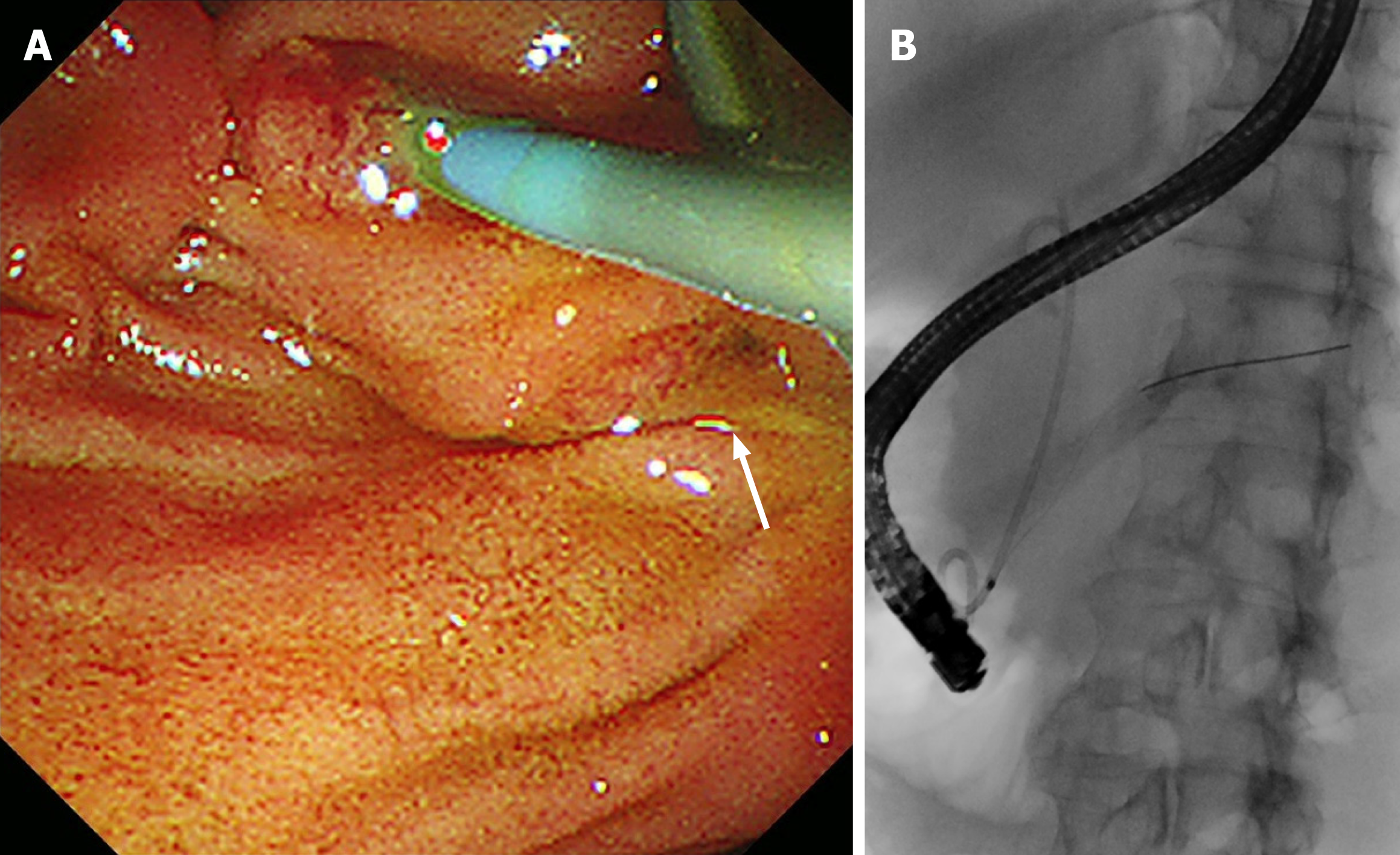
Figure 6 Second endoscopic retrograde cholangiopancreatography.
A: A separate orifice below the biliary orifice that had not been observed during the first endoscopic retrograde cholangiopancreatography is noted (white arrow); B: Selective pancreatic cannulation was easily achieved through a separate orifice.
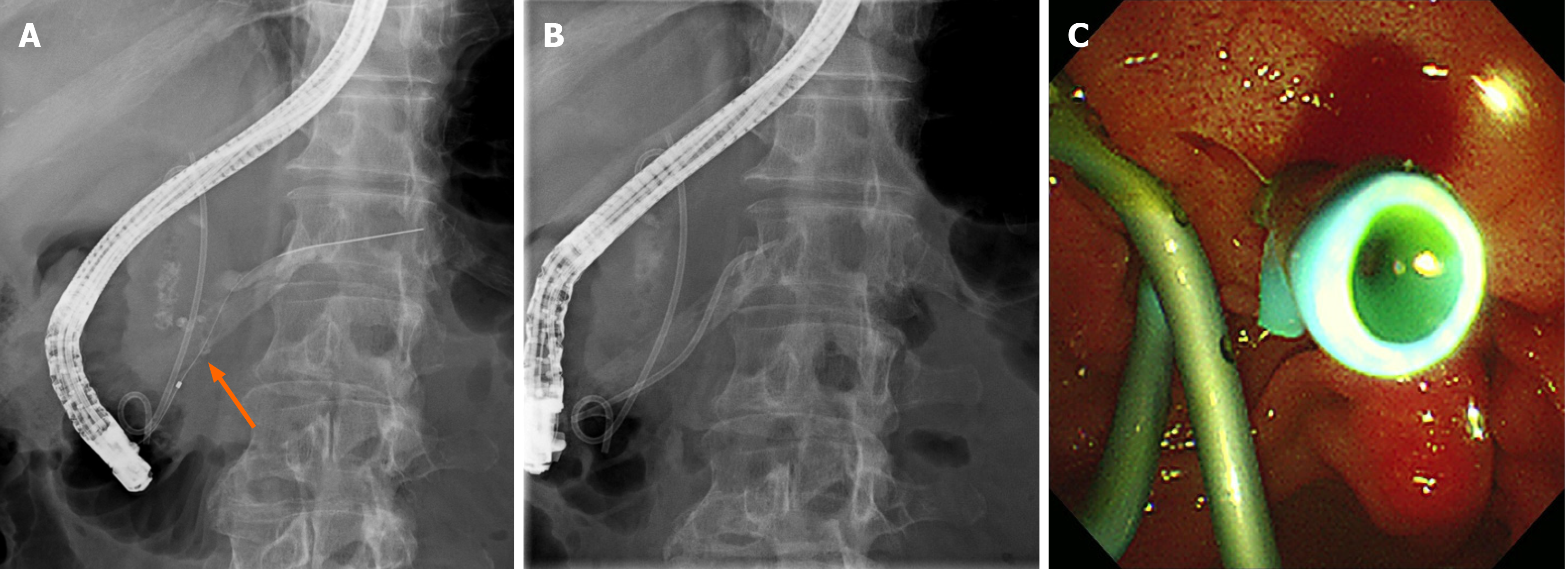
Figure 7 Second endoscopic retrograde cholangiopancreatography.
A: A pancreatogram showing dye leakage in the head of the main pancreatic duct (orange arrow); B and C: A 5F 7 cm linear plastic stent was deployed to divert the pancreatic juice.
Figure 8 Computed tomography of the abdomen two weeks after pancreatic stent placement.
Disappearance of adjacent fluid collection and edematous change of head of pancreas, and healing of the duct leakage site finding were seen.
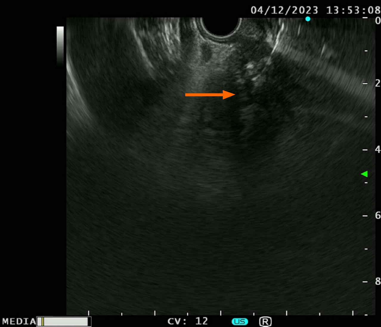
Figure 9 Retrospective analysis of endoscopic ultrasound imaging in the current case.
Suspected ductal structure within the fanning area of the target (orange arrow).
- Citation: Kim KH, Park CH, Cho E, Lee Y. Endoscopic ultrasound-guided tissue sampling induced pancreatic duct leak resolved by the placement of a pancreatic stent: A case report. World J Clin Cases 2024; 12(9): 1677-1684
- URL: https://www.wjgnet.com/2307-8960/full/v12/i9/1677.htm
- DOI: https://dx.doi.org/10.12998/wjcc.v12.i9.1677


