Copyright
©The Author(s) 2024.
World J Clin Cases. Mar 26, 2024; 12(9): 1669-1676
Published online Mar 26, 2024. doi: 10.12998/wjcc.v12.i9.1669
Published online Mar 26, 2024. doi: 10.12998/wjcc.v12.i9.1669
Figure 1 The patient developed dry asymmetric lesions on her limbs.
A: Knee joint; B: Bilateral lesser thenar; C and D: Limbs.
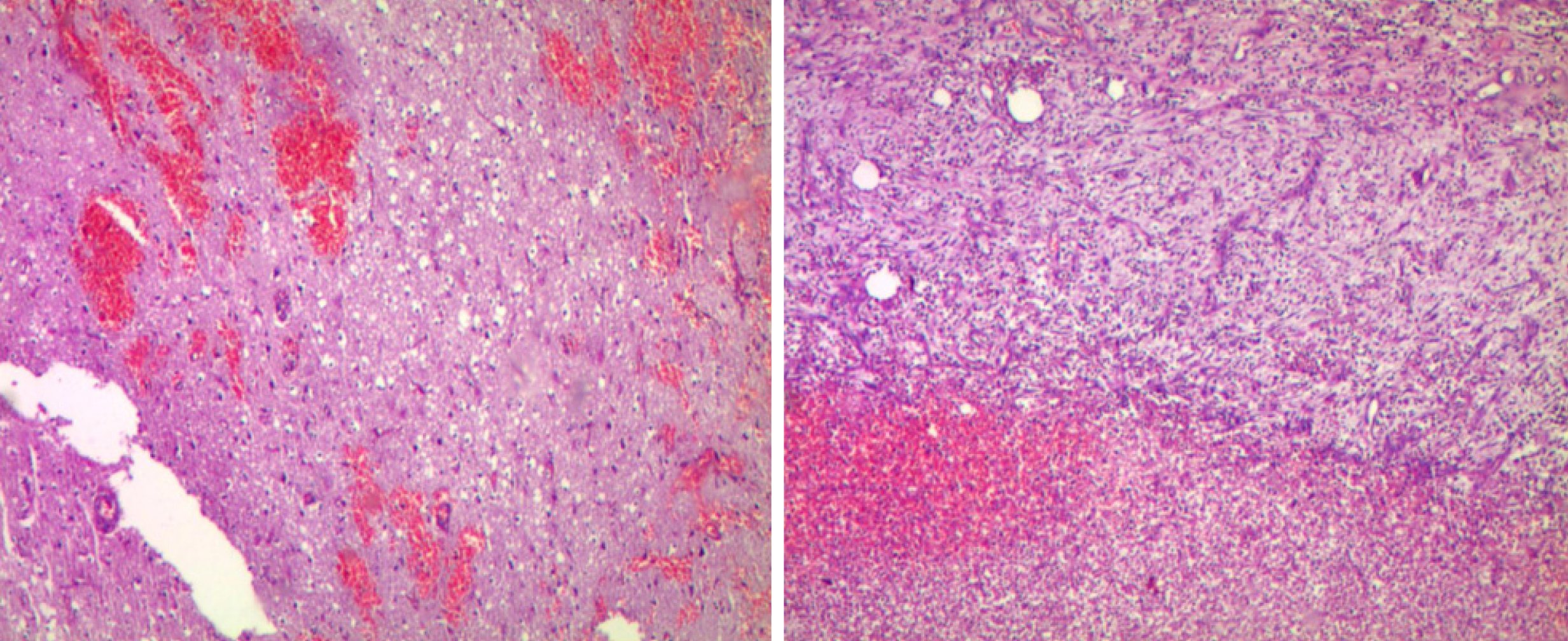
Figure 2 Pathological results of the patient’s frontal lobe brain abscess.
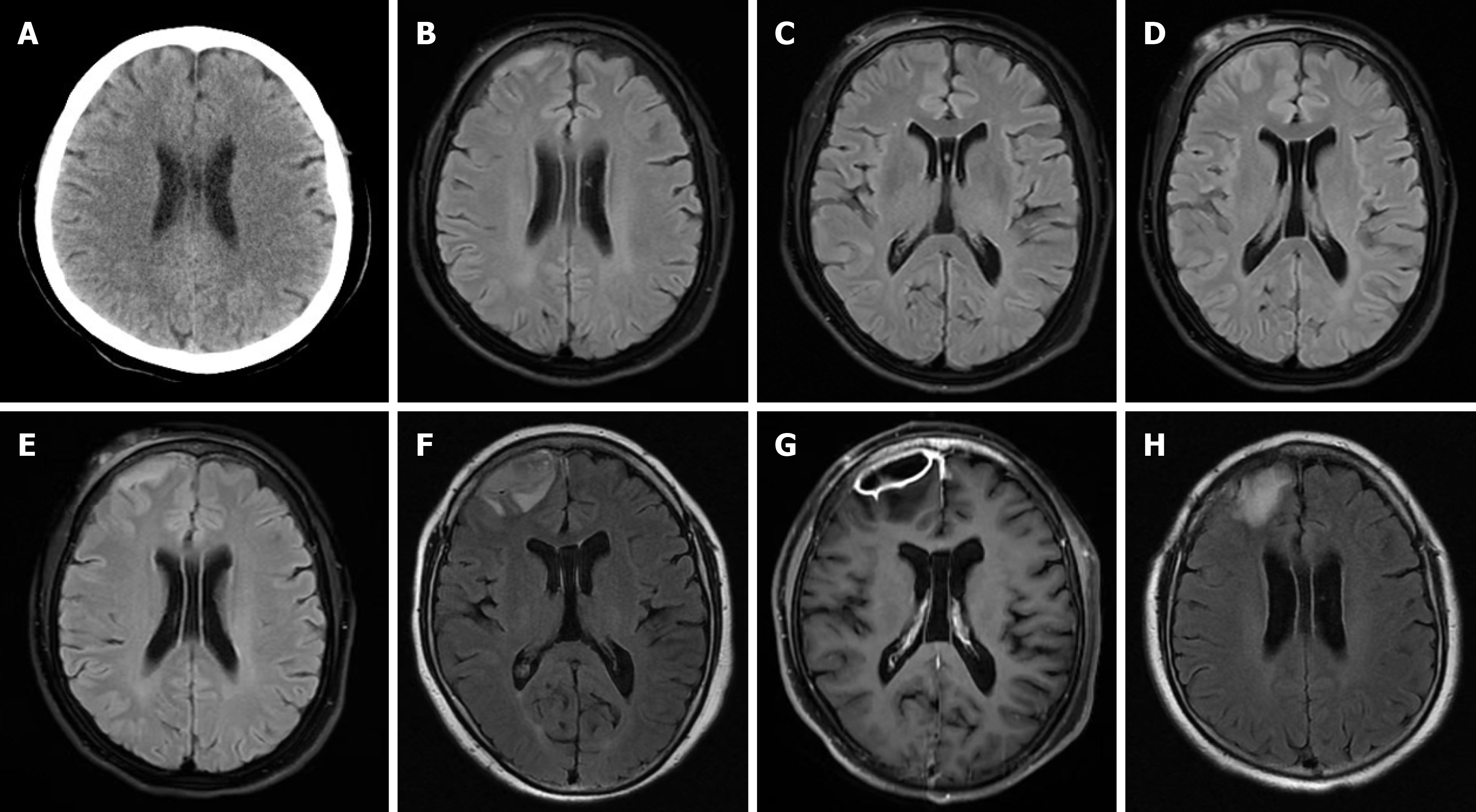
Figure 3 The patient presented radiographic features of neurological melioidosis.
A: Head computed tomography measurements were normal; B: Brain magnetic resonance imaging (MRI) suggesting subdural hematoma; C: The right frontal skin showed swelling; D and E: A second brain MRI showed that the subdural hematoma and subcutaneous swelling at the right frontal region worsened; F: The third brain MRI showed an exacerbation of the subdural lesion in the right frontal lobe, with unclear demarcation between the lesion and the right frontal lobe, suggesting an intracranial infection; G: Enhanced MRI indicated the formation of a subdural abscess in the right frontal lobe; H: Two months post-surgery for an intracranial abscess, brain MRI showed gliosis at the surgical excision site.
Figure 4 Timeline of the case.
- Citation: Ni HY, Zhang Y, Huang DH, Zhou F. Multi-systemic melioidosis in a patient with type 2 diabetes in non-endemic areas: A case report and review of literature. World J Clin Cases 2024; 12(9): 1669-1676
- URL: https://www.wjgnet.com/2307-8960/full/v12/i9/1669.htm
- DOI: https://dx.doi.org/10.12998/wjcc.v12.i9.1669


