Copyright
©The Author(s) 2024.
World J Clin Cases. Mar 16, 2024; 12(8): 1536-1543
Published online Mar 16, 2024. doi: 10.12998/wjcc.v12.i8.1536
Published online Mar 16, 2024. doi: 10.12998/wjcc.v12.i8.1536
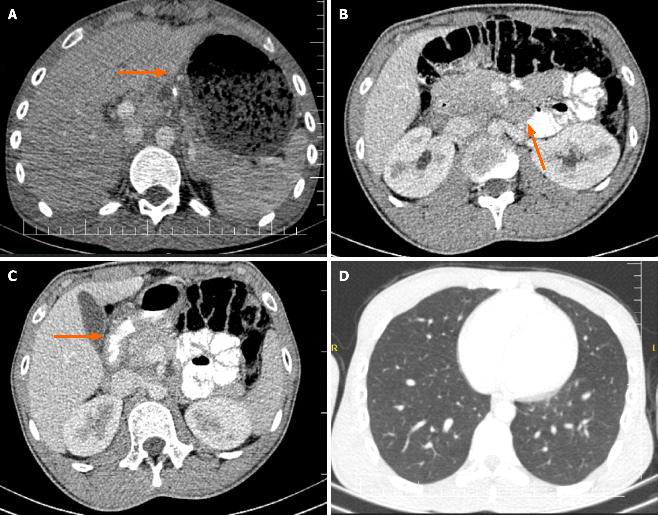
Figure 1 Oral and intravenous contrast-enhanced thoraco-abdominal computed tomography scans.
A: It demonstrates gastric distension; B: It accompanied by multiple mesenteric lymphadenopathies; C: Duodenal wall thickening and nearly complete obstruction in the second part of the duodenum; D: Thoracic computed tomography findings indicated normalcy, with no active pulmonary tuberculosis or sequelae observed.
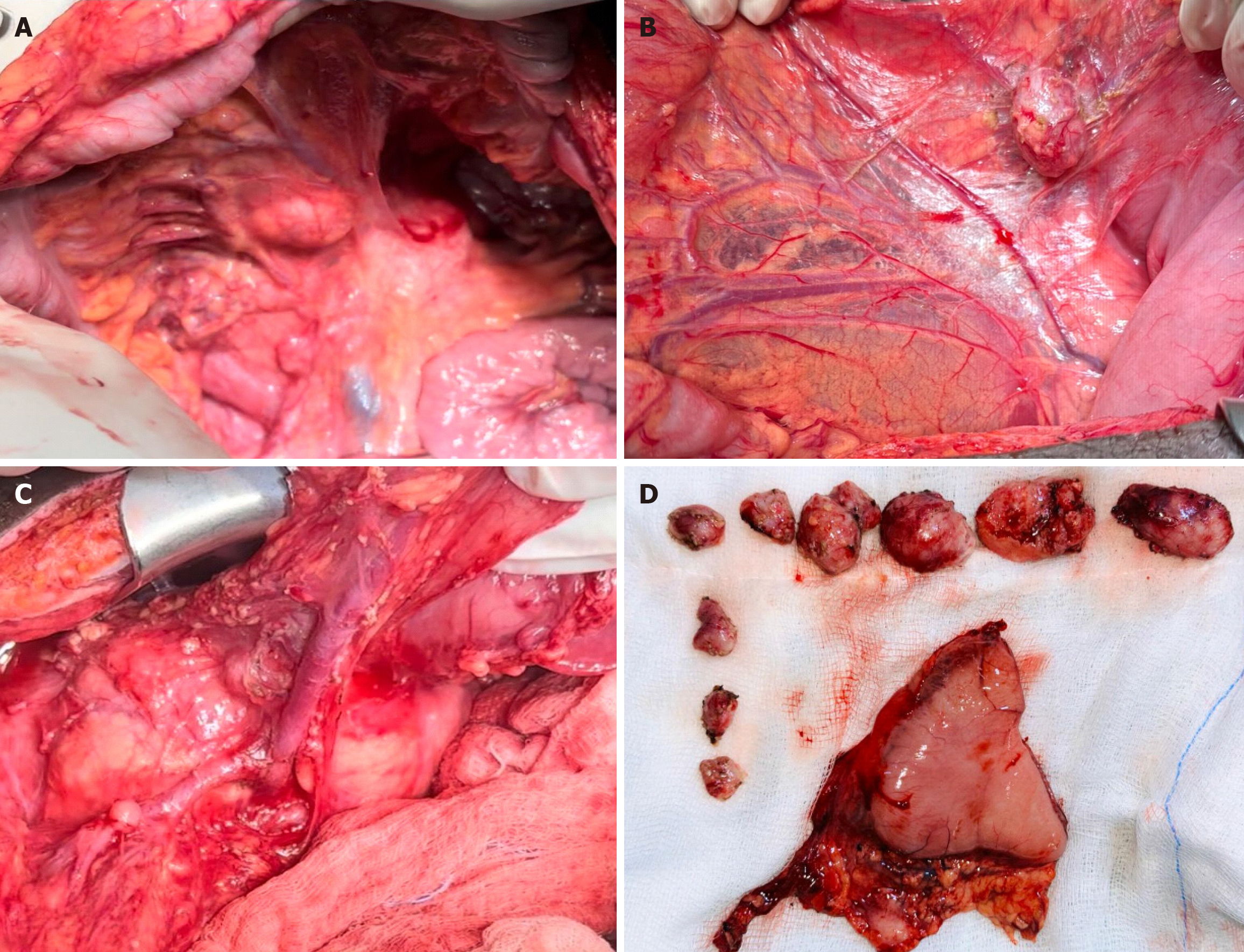
Figure 2 Intraoperative finding.
A-C: An intense inflammatory mass causing duodenal obstruction alongside multiple pathologic lymph nodes observed in the mesenteric and paraduodenal regions; D: Resected distal gastric and dissected lymph nodes.
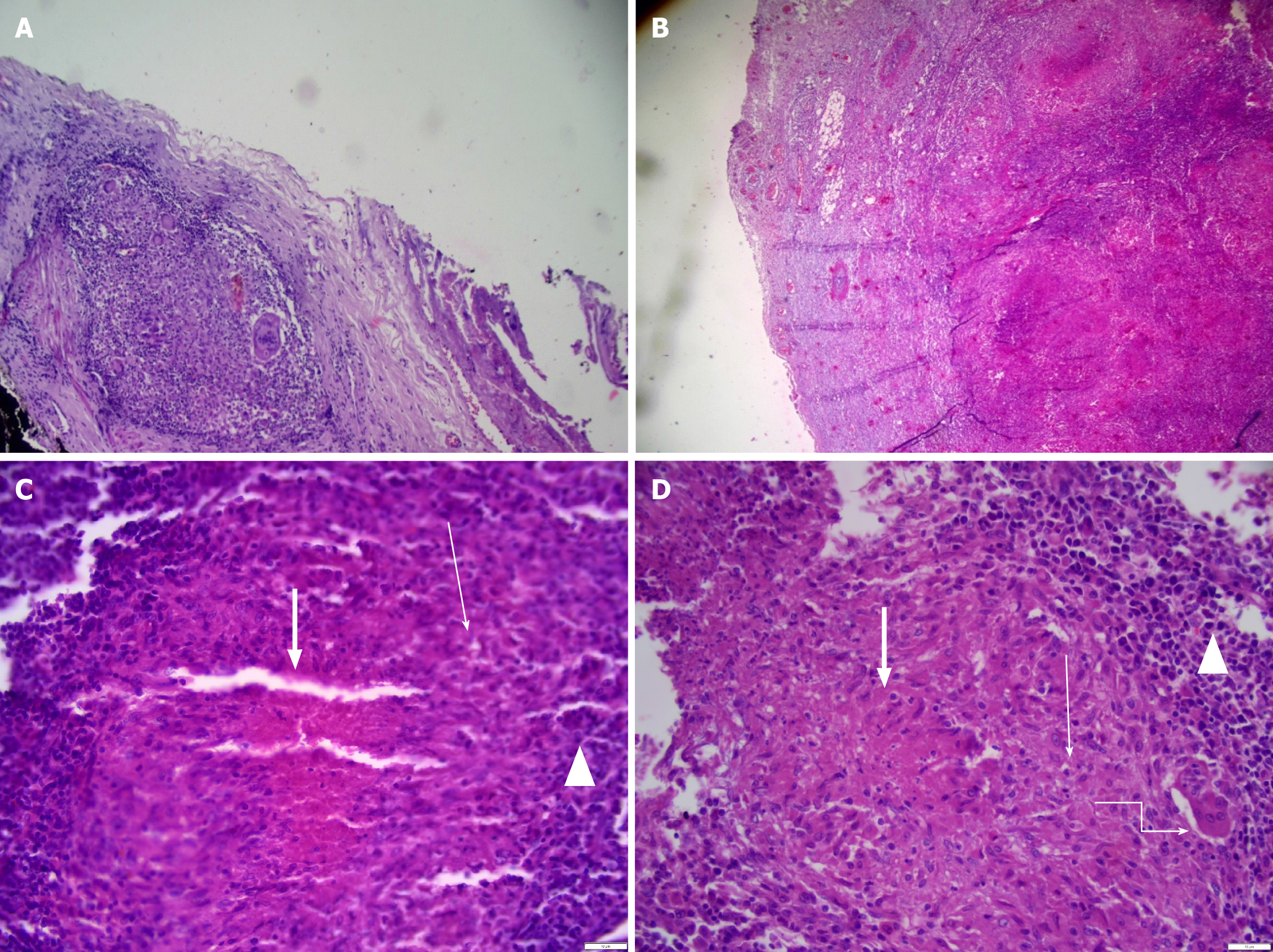
Figure 3 Histopathology.
A: Granulomatous inflammation and giant cells on the serosal surface; B: Lymph node parenchyma with effacement of multiple large tuberculoid necrotizing granuloma; C: Lymph node parenchyma with effacement of large tuberculoid necrotizing granuloma (thick arrows) epithelioid histiocytes (thin arrows) peripheral lymphocytes (triangles); D: Lymph node with tuberculoid necrotizing granuloma (thick arrows) and Langerhans giant cells (thin arrows) and peripheral lymphocytes (triangles).
- Citation: Ali AM, Mohamed YG, Mohamud AA, Mohamed AN, Ahmed MR, Abdullahi IM, Saydam T. Primary gastroduodenal tuberculosis presenting as gastric outlet obstruction: A case report and review of literature. World J Clin Cases 2024; 12(8): 1536-1543
- URL: https://www.wjgnet.com/2307-8960/full/v12/i8/1536.htm
- DOI: https://dx.doi.org/10.12998/wjcc.v12.i8.1536


