Copyright
©The Author(s) 2024.
World J Clin Cases. Mar 16, 2024; 12(8): 1504-1509
Published online Mar 16, 2024. doi: 10.12998/wjcc.v12.i8.1504
Published online Mar 16, 2024. doi: 10.12998/wjcc.v12.i8.1504
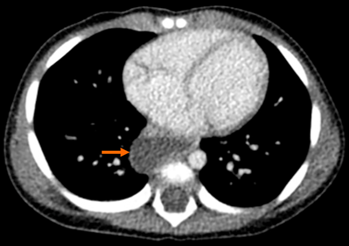
Figure 1 Computed tomography scan of the chest revealed a cystic lesion beside the oesophagus.
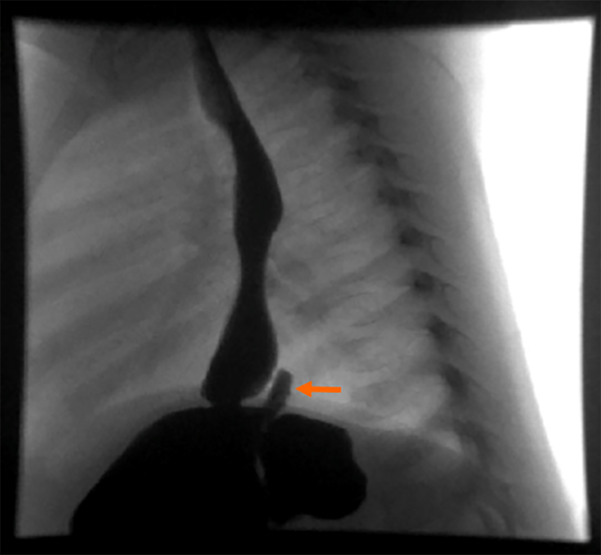
Figure 2 Contrast meal study showing a communicating gastric fundal lesion.
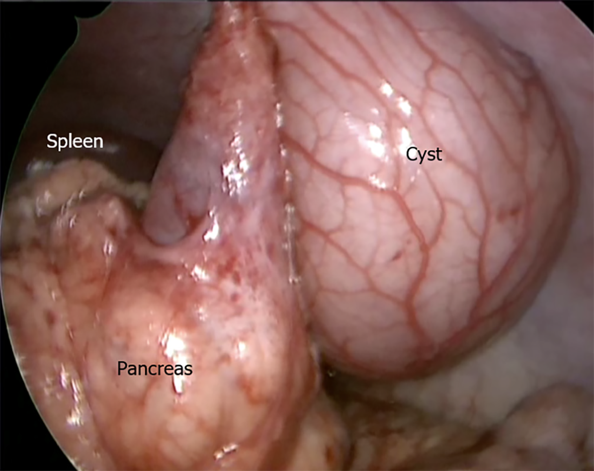
Figure 3 Intra-operative view of the pancreas with the cyst.
Figure 4 Microscopic examination confirmed the gastric origin of the duplication cysts.
A: The cyst wall consist of all layers of gastric body namely mucosa, submucosa and muscularis properia [hematoxylin and eosin (H&E) stain, original magnification ×- 40]; B: Higher power of mucosa reveals three types of cells, mucous cells, chief cells, and parietal cells (H&E stain, original magnification × 200).
- Citation: Alsinan TA, Altokhais TI. Multiple thoracic and abdominal foregut duplication cysts: A case report. World J Clin Cases 2024; 12(8): 1504-1509
- URL: https://www.wjgnet.com/2307-8960/full/v12/i8/1504.htm
- DOI: https://dx.doi.org/10.12998/wjcc.v12.i8.1504


