Copyright
©The Author(s) 2024.
World J Clin Cases. Mar 16, 2024; 12(8): 1474-1480
Published online Mar 16, 2024. doi: 10.12998/wjcc.v12.i8.1474
Published online Mar 16, 2024. doi: 10.12998/wjcc.v12.i8.1474
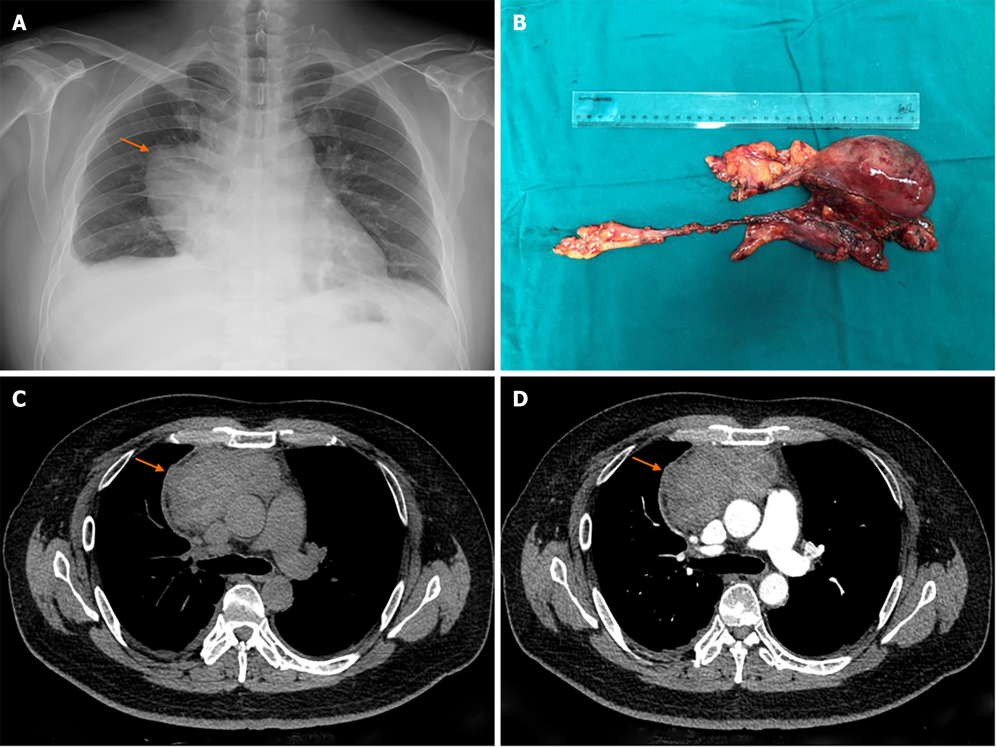
Figure 1 Radiology and macroscopic pathological image.
A: Chest X ray image; B: Macroscopic pathological image; C: Plain computer tomography (CT) image; D: CT image after contrast agent injection.
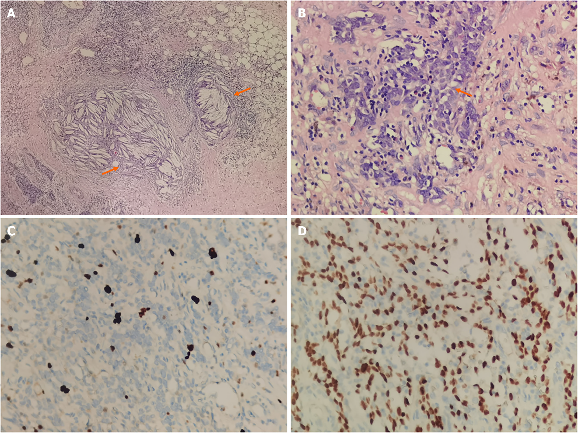
Figure 2 Histopathological picture of this case of multilocular thymic cyst.
A: The cholesterol crystals in multilocular thymic cyst (MTC); B: Squamous epithelium and lymphoid hyperplasia; C and D: Immunohistochemistry of Ki-67 and p63 in MTC. The Ki-67 was 5% positive and p63 was positive.
Figure 3 Rechecked after 9 months and no signs of recurrence.
- Citation: Sun J, Yang QN, Guo Y, Zeng P, Ma LY, Kong LW, Zhao BY, Li CM. Multilocular thymic cysts can be easily misdiagnosed as malignant tumor on computer tomography: A case report. World J Clin Cases 2024; 12(8): 1474-1480
- URL: https://www.wjgnet.com/2307-8960/full/v12/i8/1474.htm
- DOI: https://dx.doi.org/10.12998/wjcc.v12.i8.1474


