Copyright
©The Author(s) 2024.
World J Clin Cases. Mar 16, 2024; 12(8): 1467-1473
Published online Mar 16, 2024. doi: 10.12998/wjcc.v12.i8.1467
Published online Mar 16, 2024. doi: 10.12998/wjcc.v12.i8.1467
Figure 1 Imaging and macroscopic examination of malignant triton tumor in the lower abdomen.
A: Preoperative design for wide resection on the lower abdomen; B: Preoperative computed tomography (CT) depicting a 1.3 cm nodular mass in the middle of the lower abdomen; C: Preoperative positron emission tomography/CT showing mildly increased glucose metabolism at the lesion without any other significant hypermetabolic lesions.
Figure 2 Histopathologic analysis of the resected specimen.
A: Photomicrograph displaying hypocellular regions of the tumor with spindle cells, wavy nuclei, thick-walled blood vessels, perivascular accentuation of tumor cells, and some rhabdomyoblasts. Pathological biopsy revealed an MPNST with rhabdomyosarcomatous differentiation [hematoxylin and eosin (H&E staining), × 100]; B: Area showing admixture of undifferentiated small malignant cells and ovoid to round rhabdomyoblasts bearing dense hypereosinophilic cytoplasm (H&E staining, × 300).
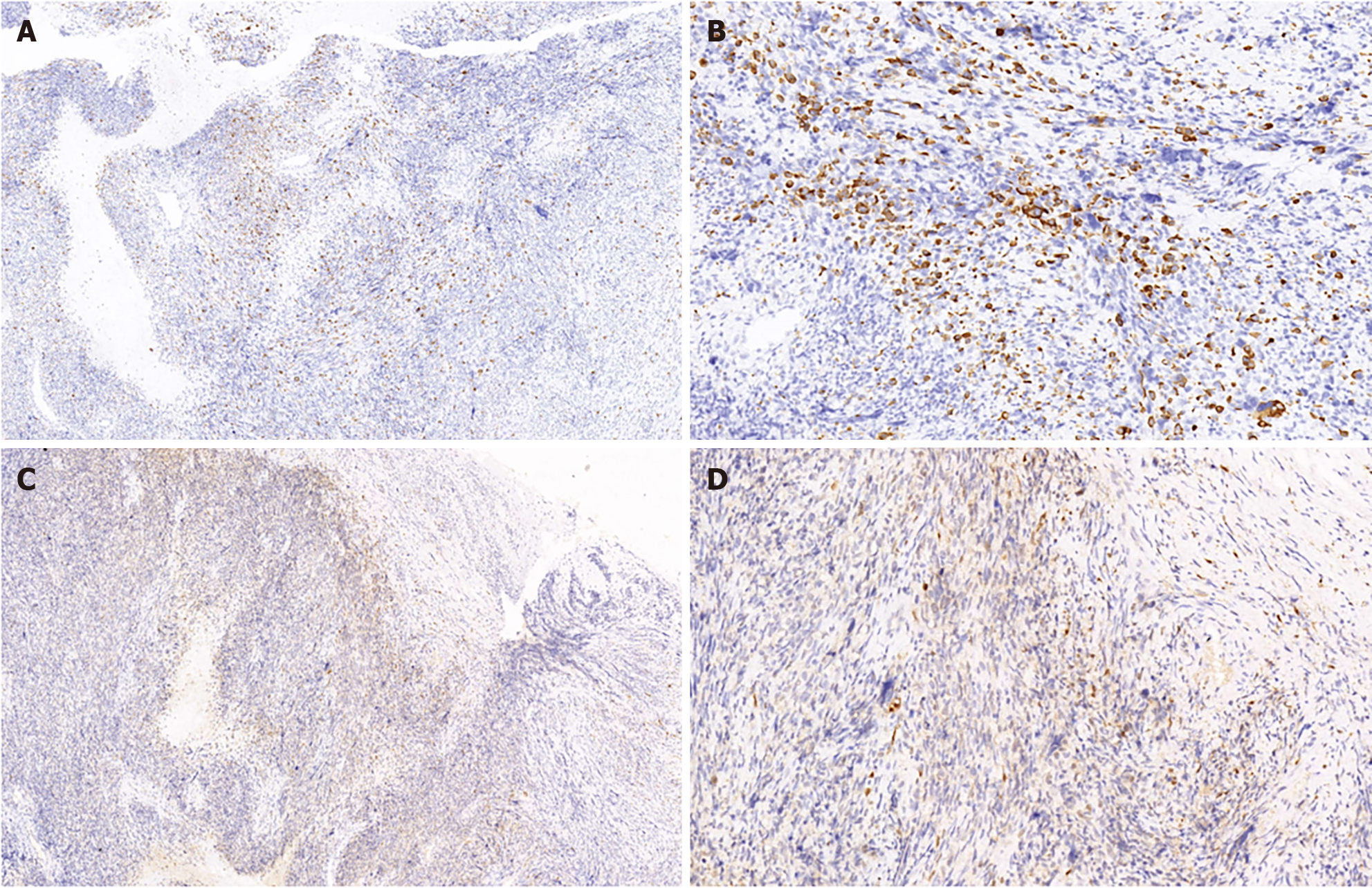
Figure 3 Immunohistochemical examination of the resected specimen.
A: Rhabdomyoblasts exhibited focal positivity for desmin (immunohistochemical staining, × 100); B: Desmin (immunohistochemical staining, × 300); C: S-100 was also detected in the malignant cells (immunohistochemical staining, × 100); D: S-100 (immunohistochemical staining, × 300).
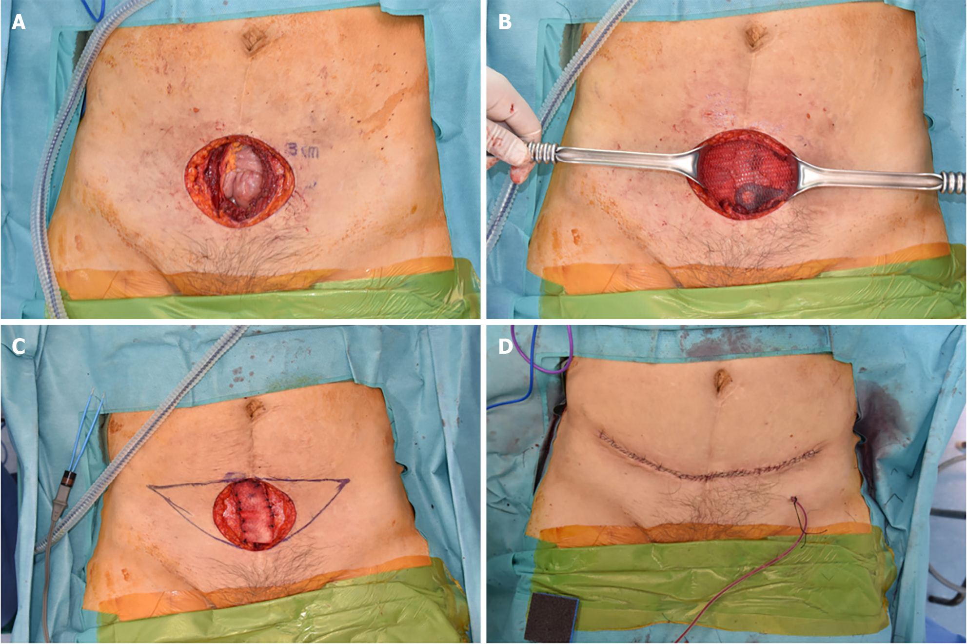
Figure 4 Surgical treatment.
A: Wide surgical excision of the mass with a 3 cm margin, inclusive of the peritoneum; B: Both sides of the peritoneum were advanced and repaired; for reinforcement, a composite mesh was inserted and affixed to both pelvic symphyses; C: The anterior fascia's deficiency was filled using an acellular dermal matrix; D: To minimize scarring, the skin defect underwent an additional excision shaped like a mini-abdominoplasty and was sutured layer-by-layer.
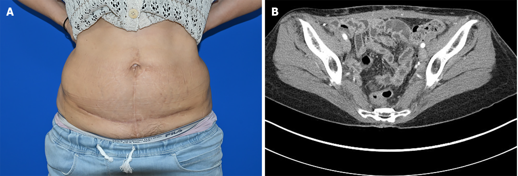
Figure 5 Follow-up.
A: Gross photo; B: Sequential follow-up abdominal computed tomography scans taken roughly every 3 mo post-resection. No instances of recurrence or complications were noted during the follow-up period.
- Citation: Yang HJ, Kim D, Lee WS, Oh SH. Malignant triton tumor in the abdominal wall: A case report. World J Clin Cases 2024; 12(8): 1467-1473
- URL: https://www.wjgnet.com/2307-8960/full/v12/i8/1467.htm
- DOI: https://dx.doi.org/10.12998/wjcc.v12.i8.1467


