Copyright
©The Author(s) 2024.
World J Clin Cases. Mar 16, 2024; 12(8): 1461-1466
Published online Mar 16, 2024. doi: 10.12998/wjcc.v12.i8.1461
Published online Mar 16, 2024. doi: 10.12998/wjcc.v12.i8.1461
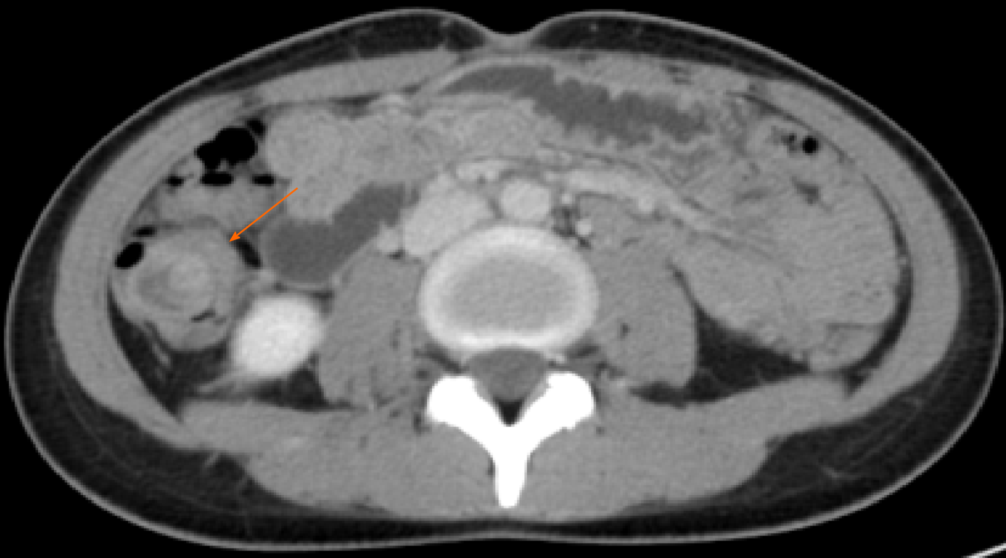
Figure 1 Preoperative computed tomography scan.
Image shows a target sign of the right colon (orange arrow).
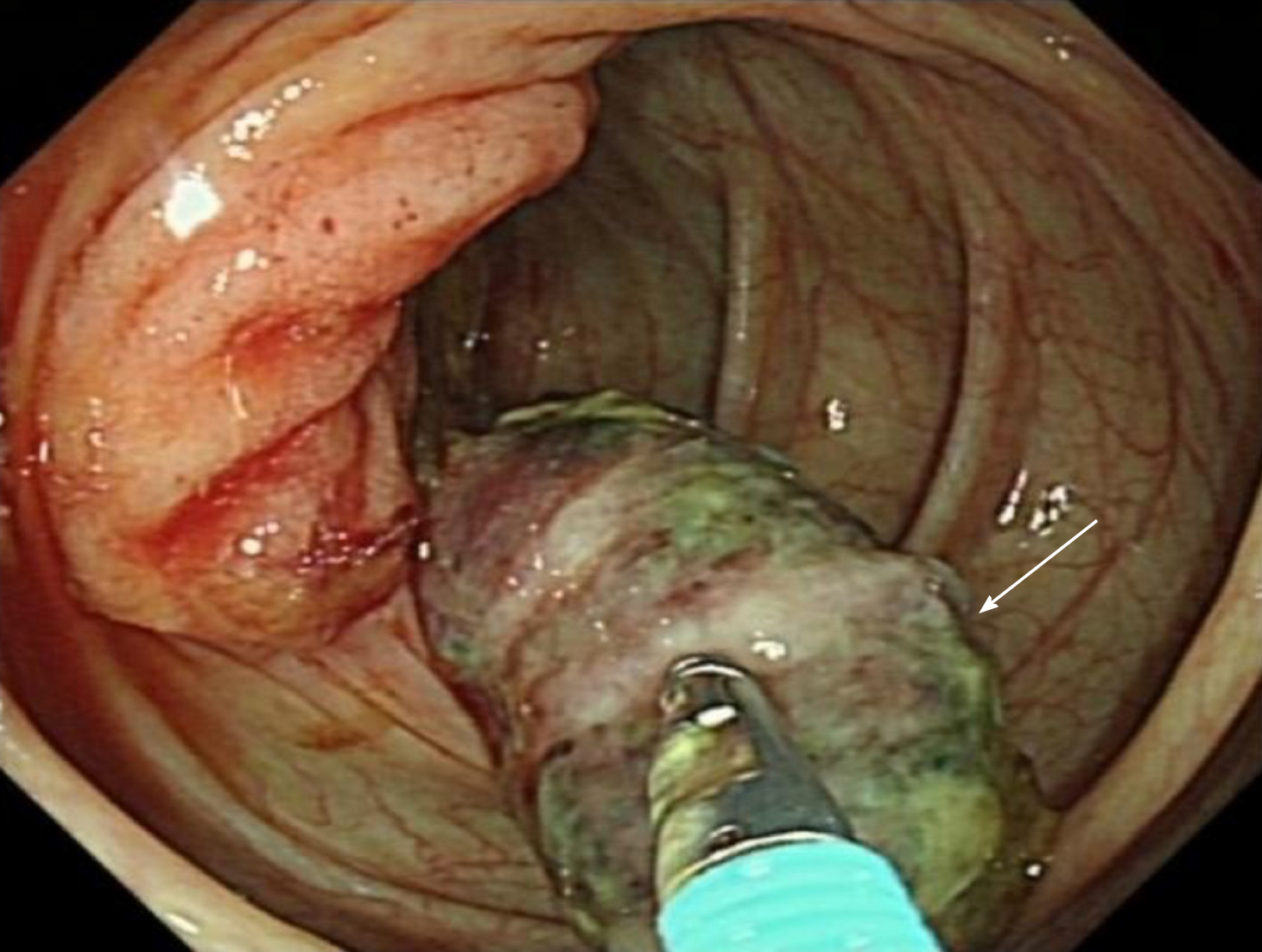
Figure 2 Colonoscopy findings.
A "finger-like" tumor is seen in the cecum with a broad head, covered with an abnormal substance, and a tendency to bleed with palpation (white arrow).
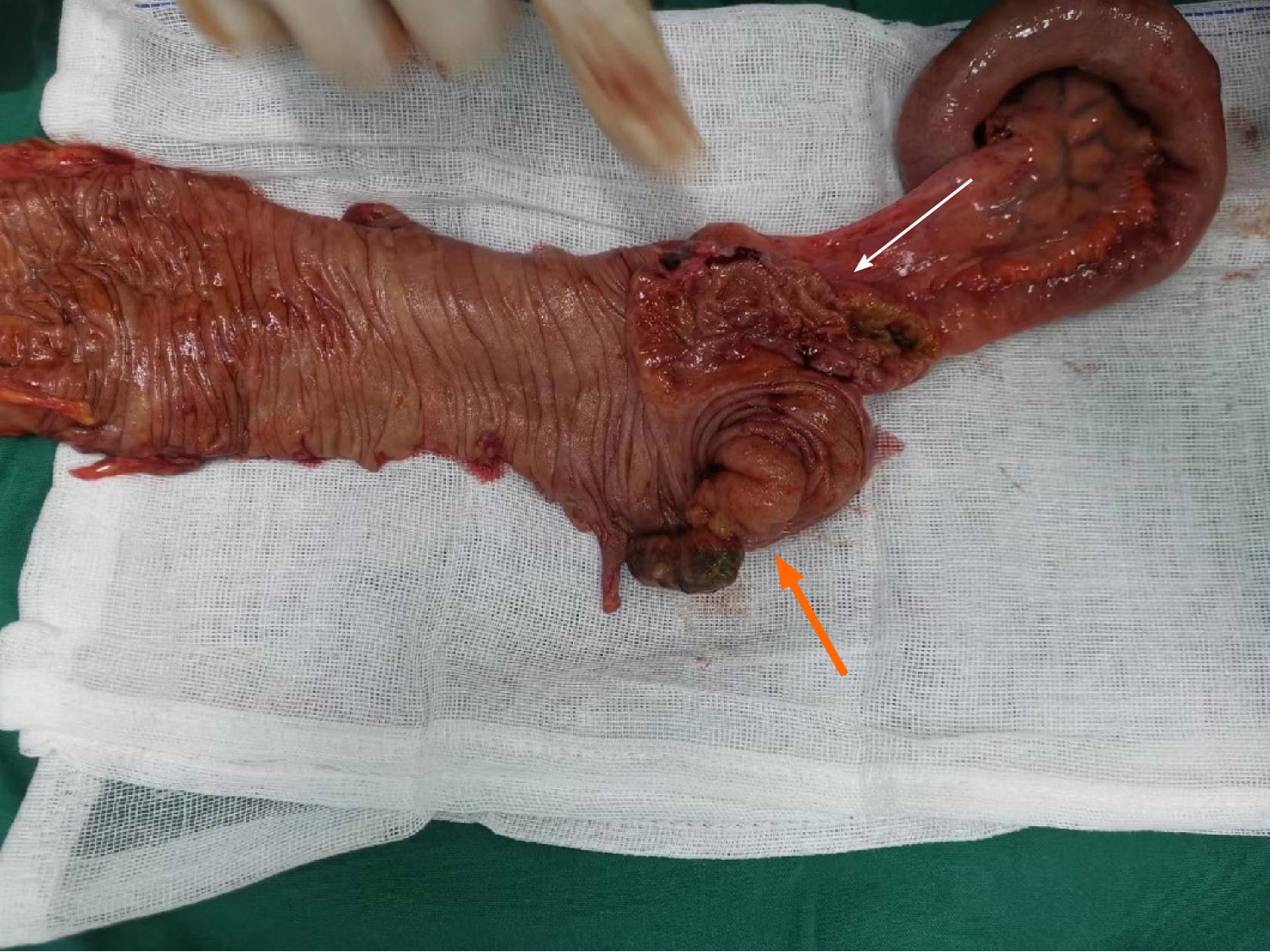
Figure 3 Colon specimen after surgery.
The white arrow indicates the tumor and the orange arrow indicates the intussusception.
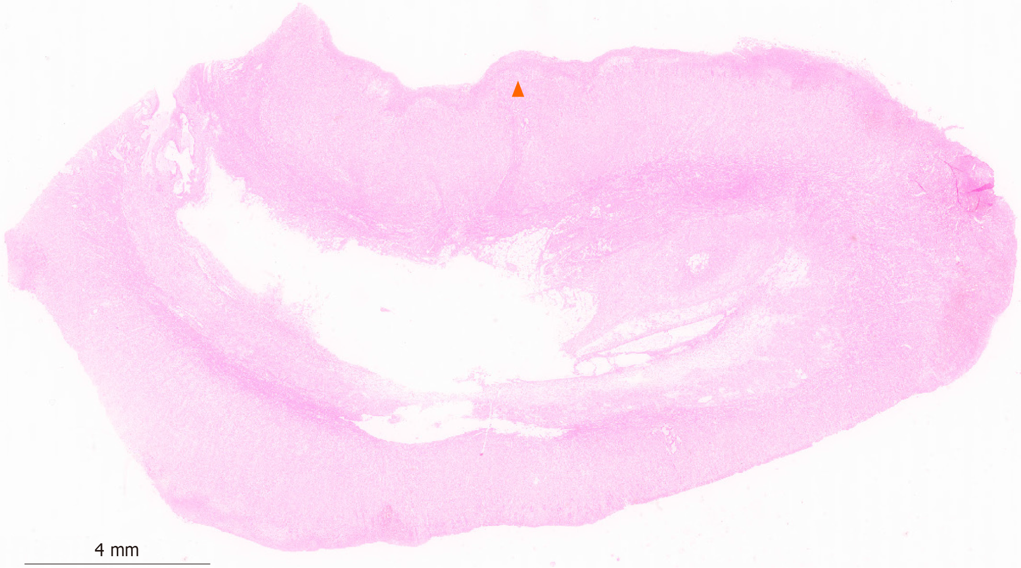
Figure 4 Tumor invasion into the muscle and subserous layers.
The lesion of the polypoid bulge in the ileocecal region is the structure of the appendix intestinal wall, and the tissue layer is the serous layer, muscle layer, submucosal layer, and mucosal layer from medial to lateral. Adenocarcinoma build-up may be seen in the muscular layer (orange triangle).
- Citation: Long Y, Xiang YN, Huang F, Xu L, Li XY, Zhen YH. Appendiceal intussusception complicated by adenocarcinoma of the cecum: A case report. World J Clin Cases 2024; 12(8): 1461-1466
- URL: https://www.wjgnet.com/2307-8960/full/v12/i8/1461.htm
- DOI: https://dx.doi.org/10.12998/wjcc.v12.i8.1461


