Copyright
©The Author(s) 2024.
World J Clin Cases. Mar 16, 2024; 12(8): 1454-1460
Published online Mar 16, 2024. doi: 10.12998/wjcc.v12.i8.1454
Published online Mar 16, 2024. doi: 10.12998/wjcc.v12.i8.1454
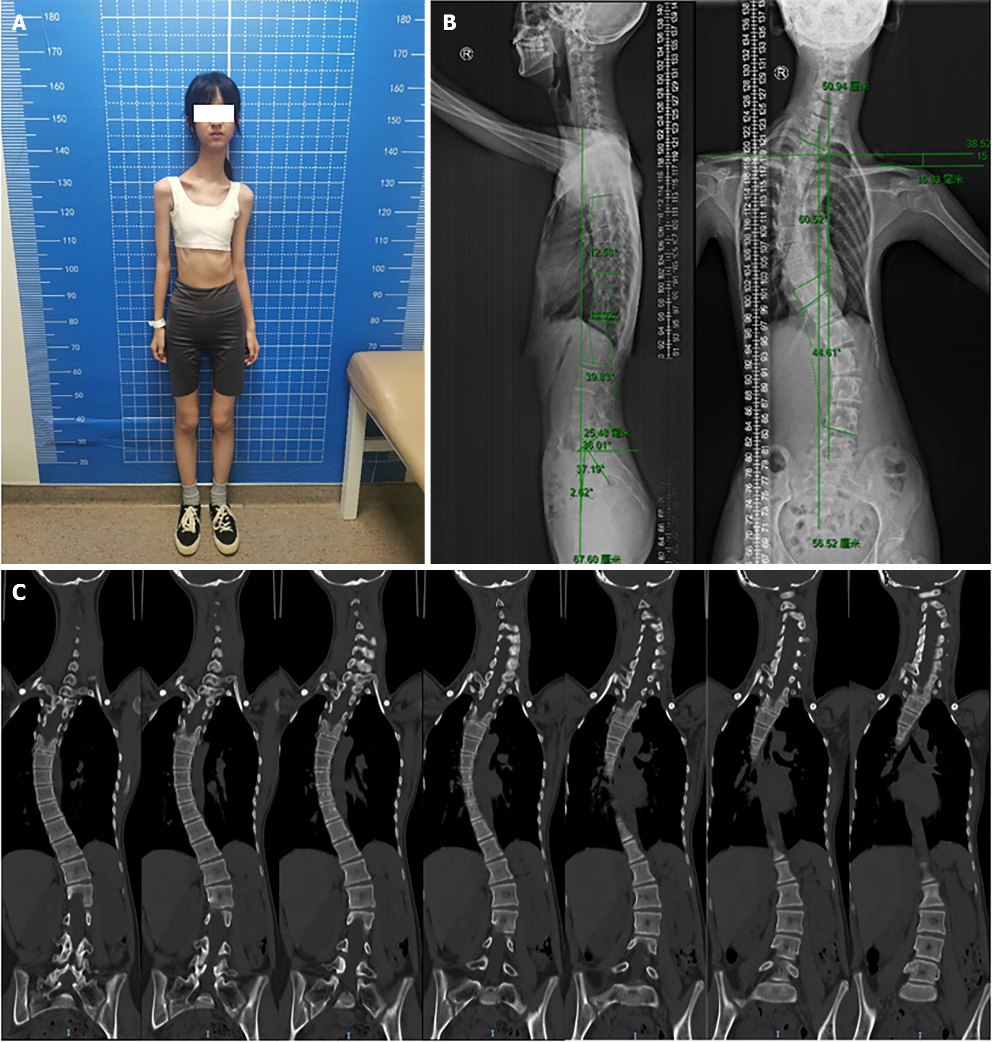
Figure 1 Clinical features of 3M syndrome.
A: Facial and body features; B: X-rays showing the slender long tubular bones, thoracic scoliosis, tall vertebral bodies and small pelvis; C: Computed tomography showing the thoracic scoliosis and tall vertebral bodies.
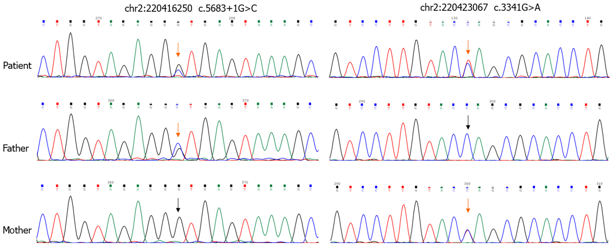
Figure 2 Schematic representation of protein and compound heterozygous variants in OBSL1.
Sequence analysis of OBSL1 showing heterozygous variant c.5683+1G>C (Splice-3) in intron and c.3341G>A (p.Trp1114Ter) in exon derived from the father and mother, respectively. Father were heterozygous for the c.5683+1G>C variant and mother were heterozygous for the c.3341G>A variant.
- Citation: Luo MR, Dai SM, Li Y, Wang Q, Liu H, Gao P, Liu JY, Chen J, Zhao SJ, Yin GY. 3M syndrome patient with a novel mutation: A case report. World J Clin Cases 2024; 12(8): 1454-1460
- URL: https://www.wjgnet.com/2307-8960/full/v12/i8/1454.htm
- DOI: https://dx.doi.org/10.12998/wjcc.v12.i8.1454


