Copyright
©The Author(s) 2024.
World J Clin Cases. Mar 16, 2024; 12(8): 1430-1436
Published online Mar 16, 2024. doi: 10.12998/wjcc.v12.i8.1430
Published online Mar 16, 2024. doi: 10.12998/wjcc.v12.i8.1430
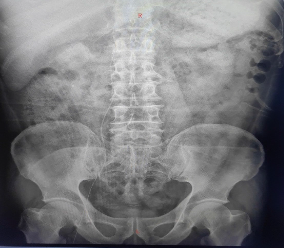
Figure 1 Catheter tip at the lumbar 1 segment.
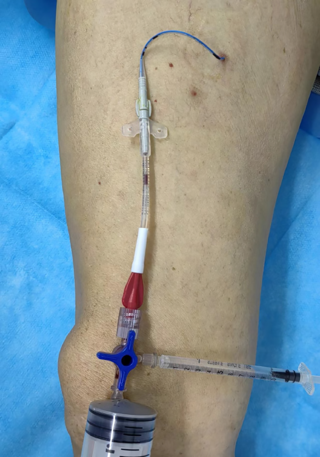
Figure 2 Coagulated blood in the catheter and extension tube.
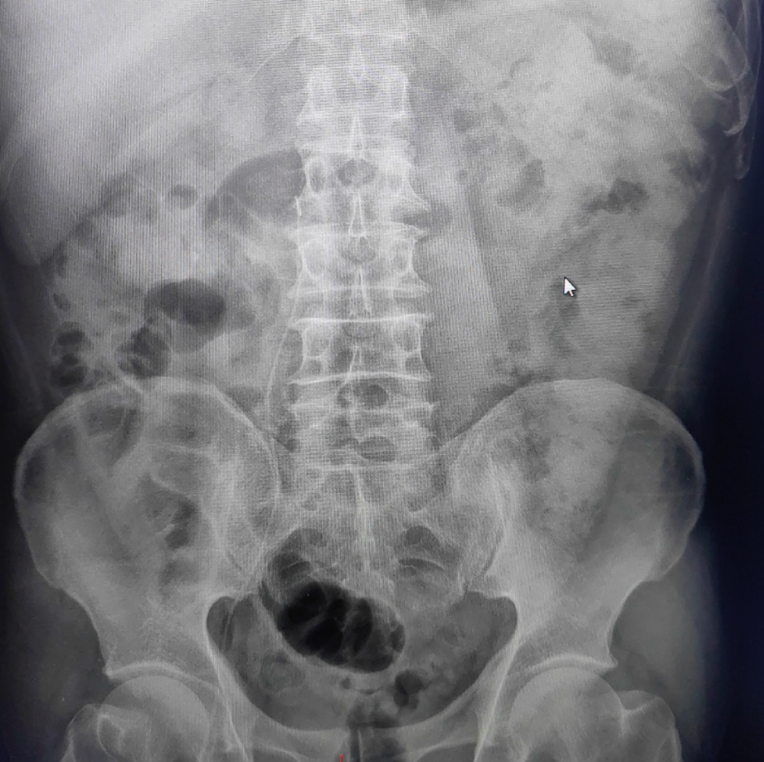
Figure 3 The bent catheter tip at the lumbar 4 segment.
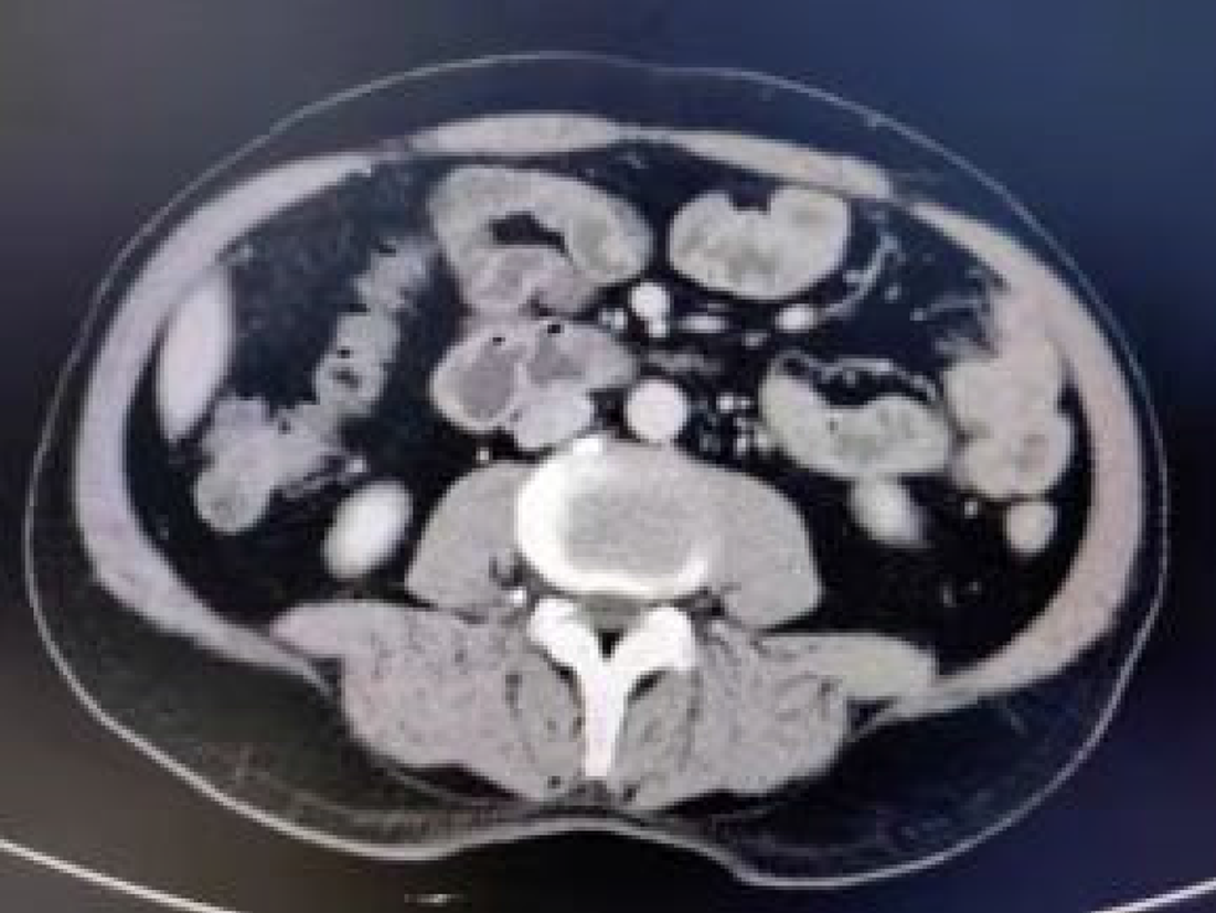
Figure 4 Computed tomography image revealing that the catheter was not in the superior vena cava.
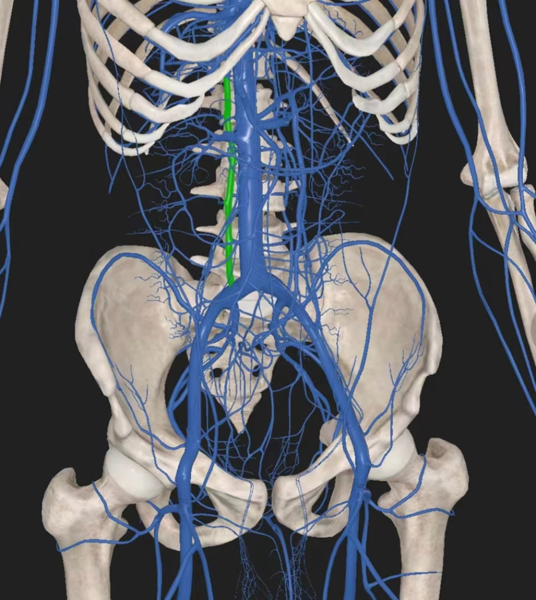
Figure 5 Three-dimensional body vascular anatomy.
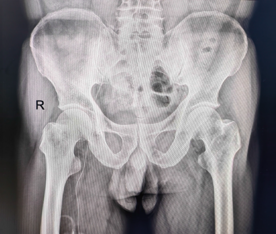
Figure 6 Catheter tip in the external iliac vein.
- Citation: Zhu XJ, Zhao L, Peng N, Luo JM, Liu SX. Lower extremity peripherally inserted central catheter placement ectopic to the ascending lumbar vein: A case report. World J Clin Cases 2024; 12(8): 1430-1436
- URL: https://www.wjgnet.com/2307-8960/full/v12/i8/1430.htm
- DOI: https://dx.doi.org/10.12998/wjcc.v12.i8.1430


