Copyright
©The Author(s) 2024.
World J Clin Cases. Mar 16, 2024; 12(8): 1422-1429
Published online Mar 16, 2024. doi: 10.12998/wjcc.v12.i8.1422
Published online Mar 16, 2024. doi: 10.12998/wjcc.v12.i8.1422
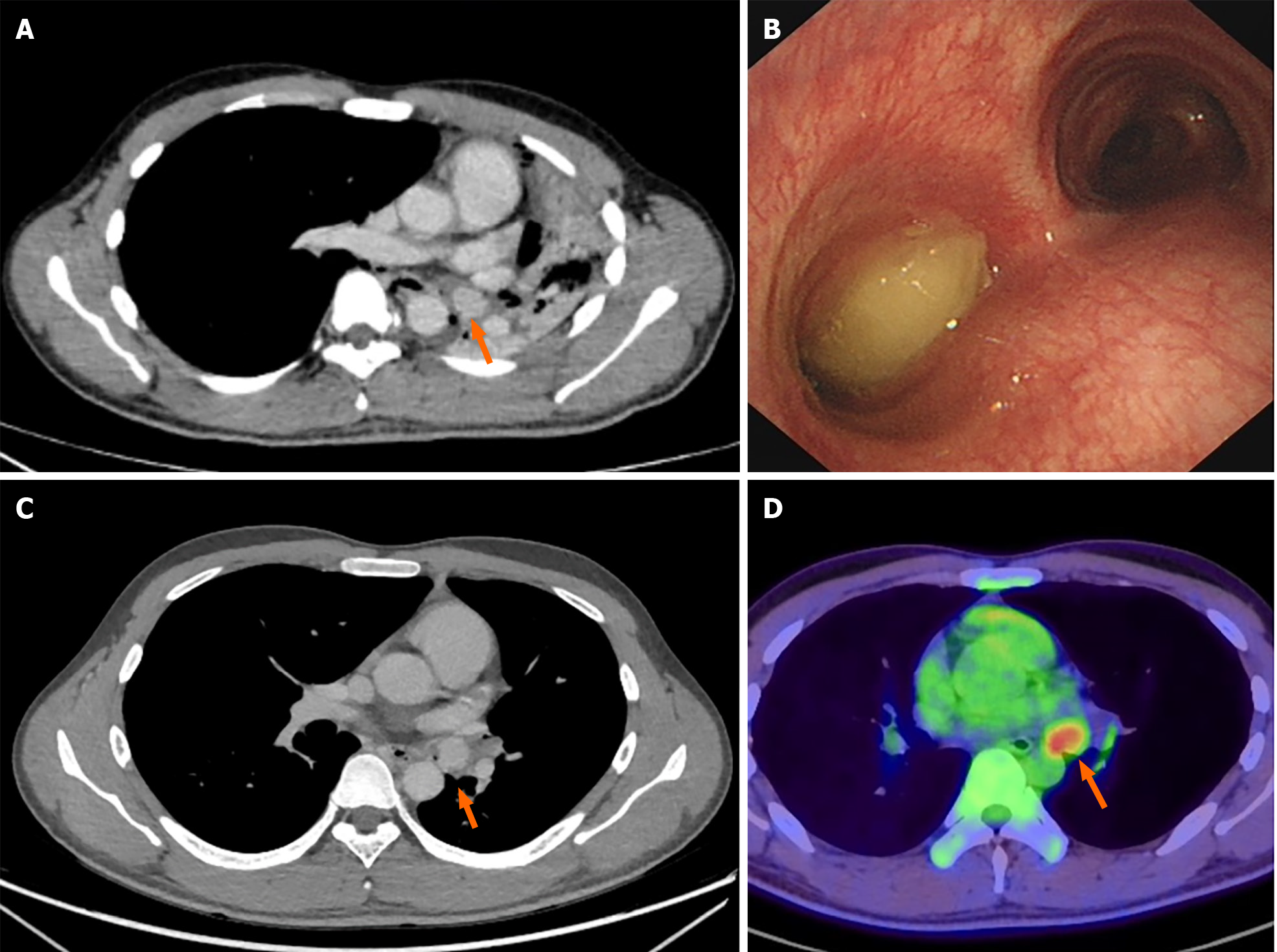
Figure 1 Preoperative patient examination.
A: Chest computed tomography (CT) scan with enhanced imaging revealed a 2-cm soft tissue density nodule situated in the left main bronchus, resulting in complete atelectasis of the left lung; B: Electronic bronchoscopy image showed a polypoid tumor obstructing the left main bronchus, featuring a surface covered with gray yellow necrotic material; C: CT imaging demonstrated signs of recruitment in the left upper lung after endoscopic treatment, while the mass in the left main bronchus persisted; D: Whole-body positron emission tomography/CT scan revealed metabolically active lesions indicative of malignancy, with the tumor location highlighted by a red arrow.
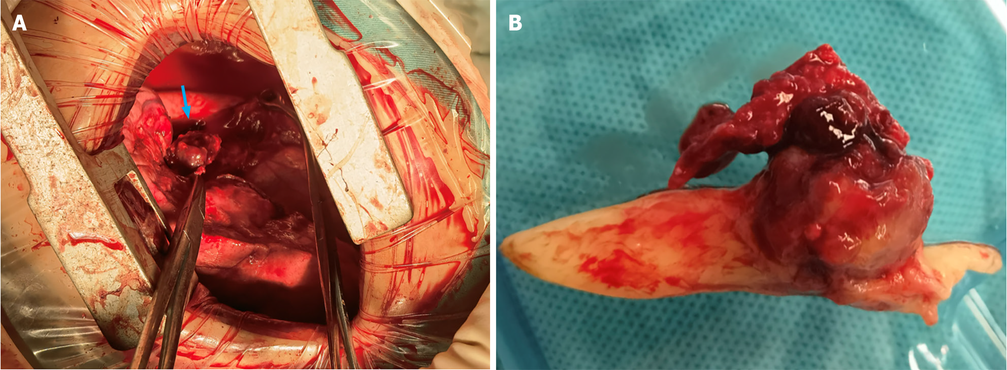
Figure 2 Intraoperative images.
A: An anterolateral incision facilitated sleeve resection of the left lower lobe and systematic mediastinal lymph node dissection. The image depicts the hyperinflated left upper lung, the severed left lower lobe bronchus, and a 2 cm dark red spherical tumor obstructing the bronchial opening in the left lower lobe while infiltrating the bronchial wall (indicated by a blue arrow); B: The tumor was accompanied by a white, tongue-shaped, clotted sputum plug extending into the left upper bronchus and obstructing a substantial portion of the lumen.
Figure 3 Gross and microscopic hematoxylin-eosin staining examination of the mass.
A: The mass, measuring 2.5 cm × 2 cm × 1 cm, was well circumscribed and displays a gray white color after formalin fixation; B: The mass was predominantly composed of clear cells with abundant clear or slightly eosinophilic cytoplasm and small round nuclei. The mass contains variable amounts of amorphous eosinophilic material interspersed between clear cells; C: Additionally, some areas of the neoplasm feature foci of mucus-secreting cells were intermixed with clear cells.
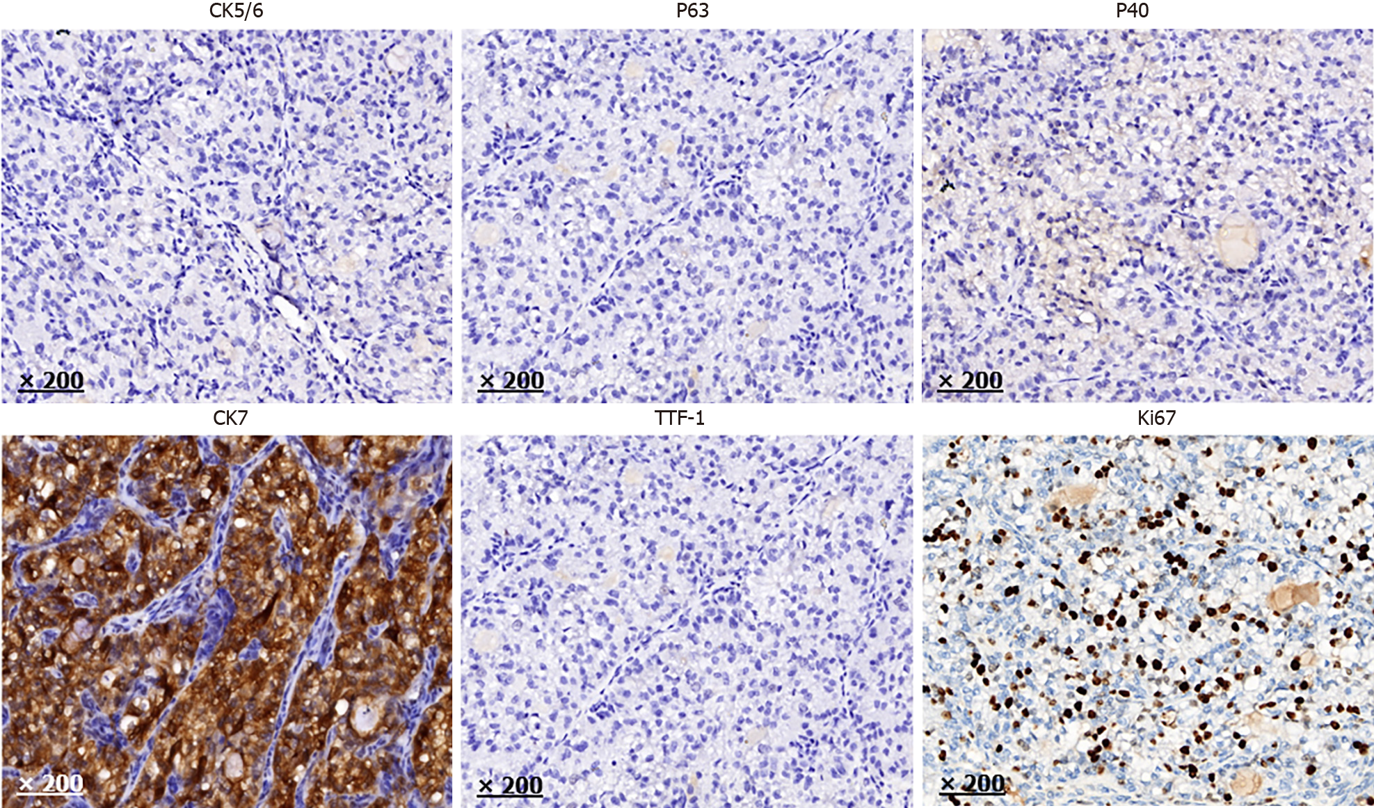
Figure 4 Immunohistochemistry.
The tumor cells showed diffuse positivity for cytokeratin (CK) 7 and negativity for cytokeratin 5/6, P63, P40, and thyroid transcription factor-1. The Kiel 67 proliferation index of tumor cells in hotspots were relatively high. CK: Cytokeratin; TTF-1: Thyroid transcription factor-1.
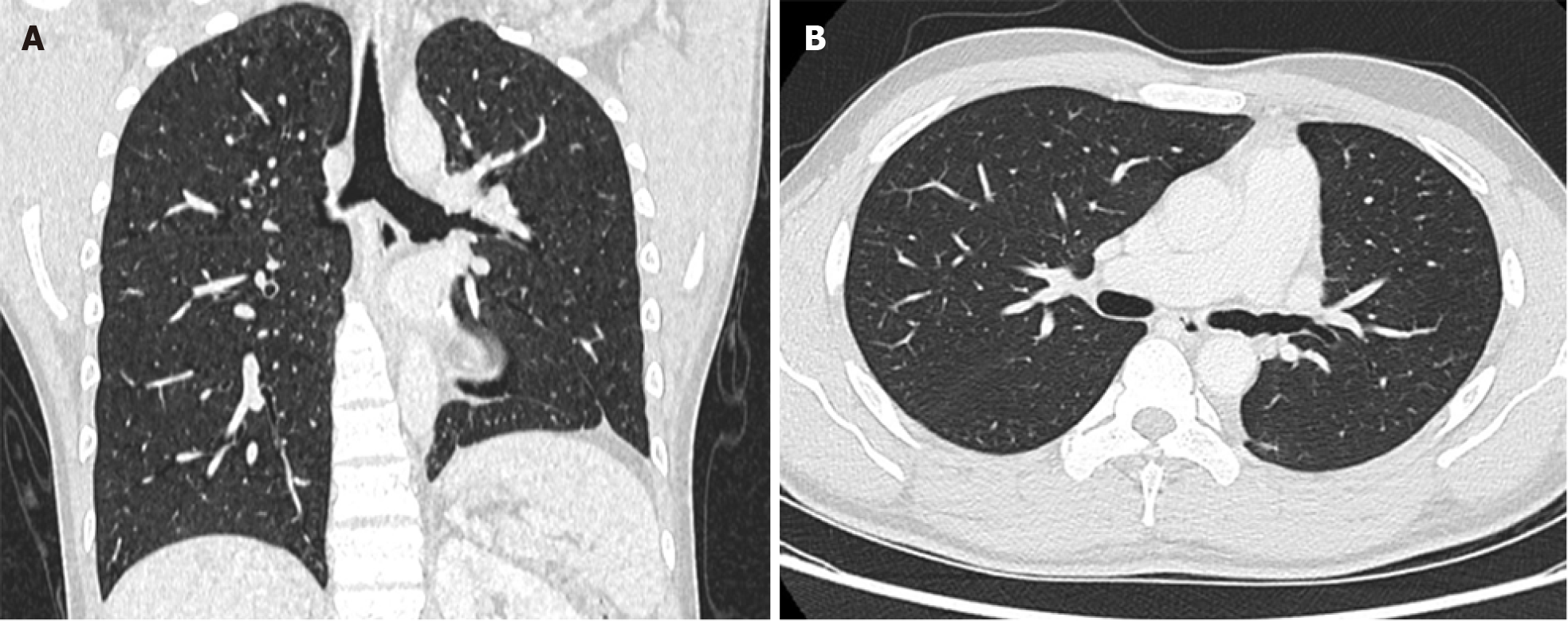
Figure 5 High-resolution chest contrast-enhanced computed tomography.
A: Coronal plane; B: Transverse plane. A re-examination via computed tomography 1.5 yrs after the operation revealed that the lungs were in good condition, with no evidence of recurrence.
- Citation: Yu XH, Wang WX, Yang DS, Gong LH. Left lower lobe sleeve resection for the clear cell variant of pulmonary mucoepidermoid carcinoma: A case report. World J Clin Cases 2024; 12(8): 1422-1429
- URL: https://www.wjgnet.com/2307-8960/full/v12/i8/1422.htm
- DOI: https://dx.doi.org/10.12998/wjcc.v12.i8.1422


