Copyright
©The Author(s) 2024.
World J Clin Cases. Mar 6, 2024; 12(7): 1356-1364
Published online Mar 6, 2024. doi: 10.12998/wjcc.v12.i7.1356
Published online Mar 6, 2024. doi: 10.12998/wjcc.v12.i7.1356
Figure 1 Preoperative magnetic resonance imaging image of the cervical spine showing C3-C7 spinal canal stenosis with spinal cord edema.
A: Sagittal T2 weighted imaging (T2WI); B: Sagittal T2WI + fat suppression; C and D: Axial T2WI at the C5/6 (C) and C6/7 (D) levels showing traumatic disc rupture, protrusion and spinal cord compression.
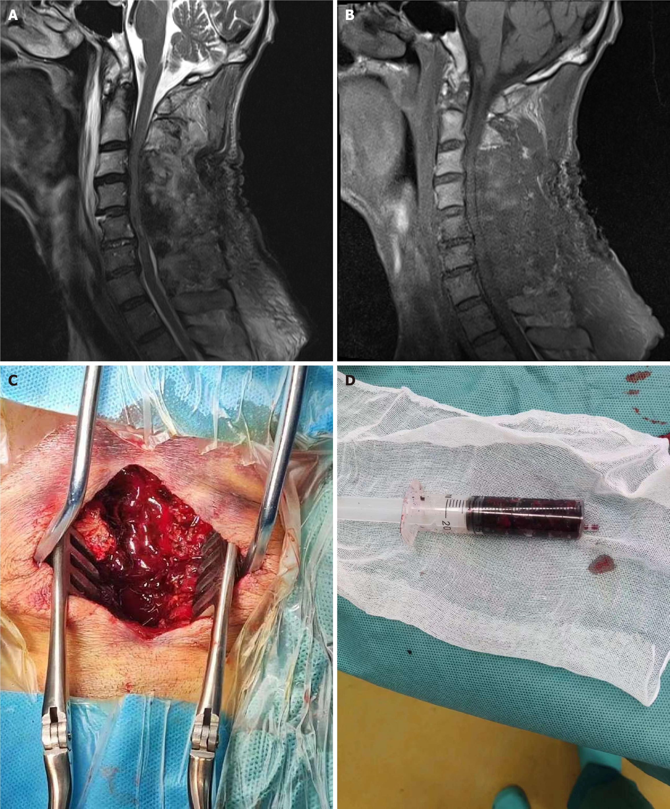
Figure 2 Postoperative magnetic resonance imaging obtained 8 h after symptom onset and the intraoperative view.
A: Magnetic resonance imaging (MRI) showing heterogeneous intensity on T2 weighted imaging (T2WI); B: MRI showing an isointense low signal intensity on T1WI with marked indentation on the dural sac; C and D: Intraoperative image showing the spinal epidural hematoma.
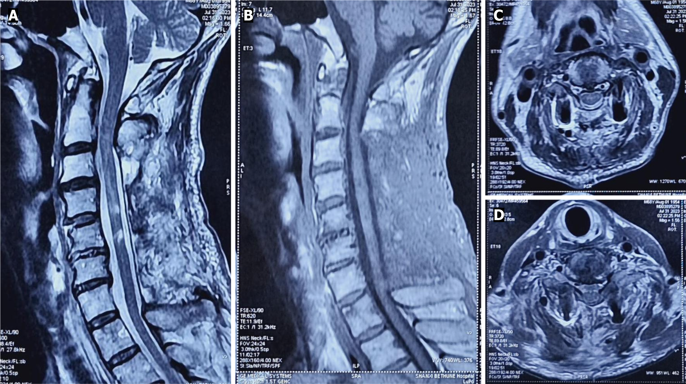
Figure 3 Postoperative magnetic resonance imaging taken 30 d after epidural hematoma elimination.
A: Sagittal T2 weighted imaging (T2WI); B: Sagittal T1WI; C and D: Axial T2WI at the C5/6 (C) and C6/7 (D) levels. Epidural hematoma has completely vanished, and the central spinal cord showed high signal at the level of C4-C5 on T2WI.
- Citation: Yan RZ, Chen C, Lin CR, Wei YH, Guo ZJ, Li YK, Zhang Q, Shen HY, Sun HL. Delayed neurological dysfunction following posterior laminectomy with lateral mass screw fixation: A case report and review of literature. World J Clin Cases 2024; 12(7): 1356-1364
- URL: https://www.wjgnet.com/2307-8960/full/v12/i7/1356.htm
- DOI: https://dx.doi.org/10.12998/wjcc.v12.i7.1356


