Copyright
©The Author(s) 2024.
World J Clin Cases. Mar 6, 2024; 12(7): 1346-1355
Published online Mar 6, 2024. doi: 10.12998/wjcc.v12.i7.1346
Published online Mar 6, 2024. doi: 10.12998/wjcc.v12.i7.1346
Figure 1 Preoperative clinical view.
An increase in volume corresponding to the circumscribed, well delimited lesion, between 2.2 and 2.3.
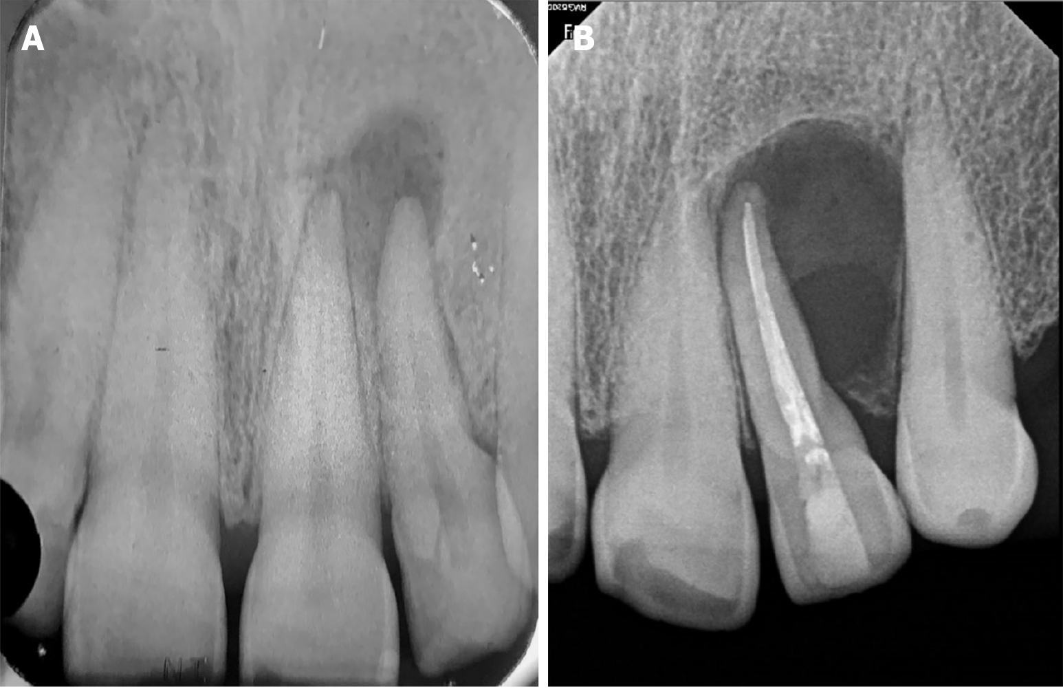
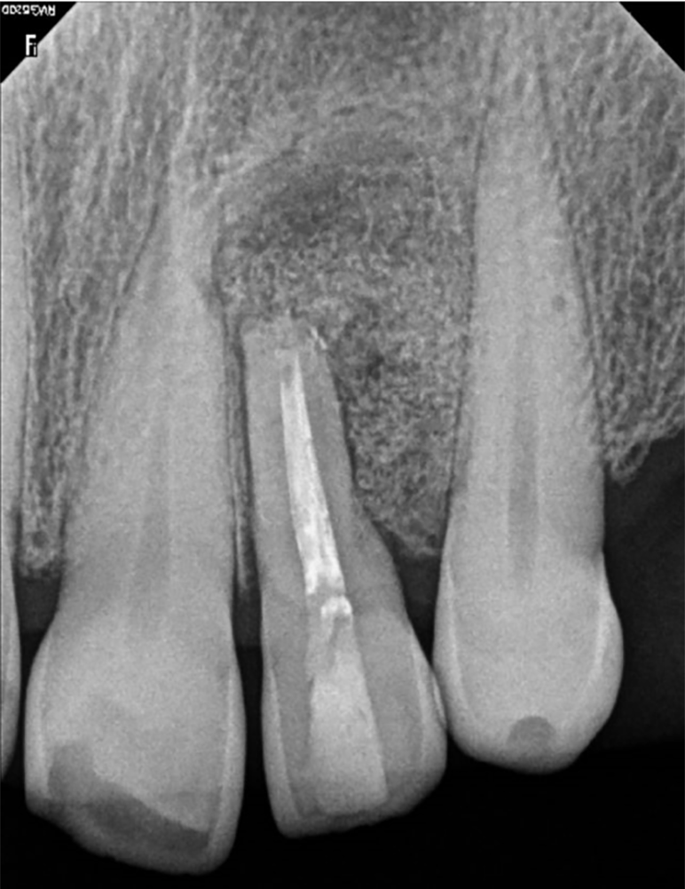
Figure 2 Preoperative periapical radiograph.
A: X-ray prior to root canal treatment, 6 years ago; B: Current initial radiograph showing a well-defined and circumscribed radiolucent area at the apex of tooth 22.
Figure 3 Cone-beam computed tomography view.
A: Sagittal; B: Axial; C: Coronal.
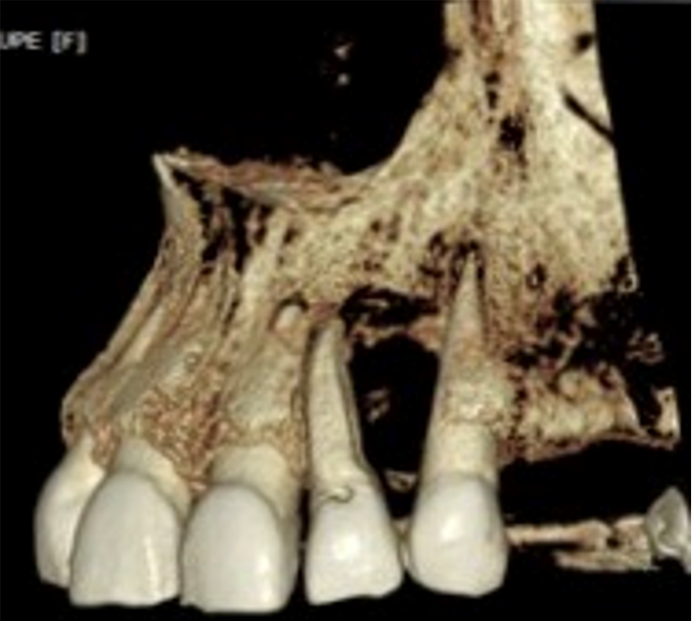
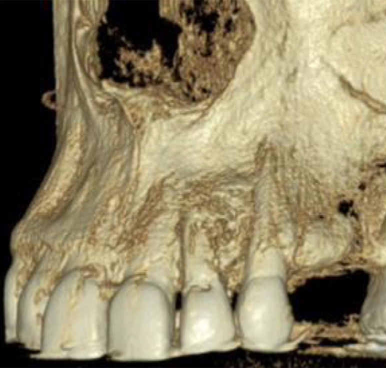
Figure 4 Three-dimensional model reconstruction.
Note the absence of vestibular wall.
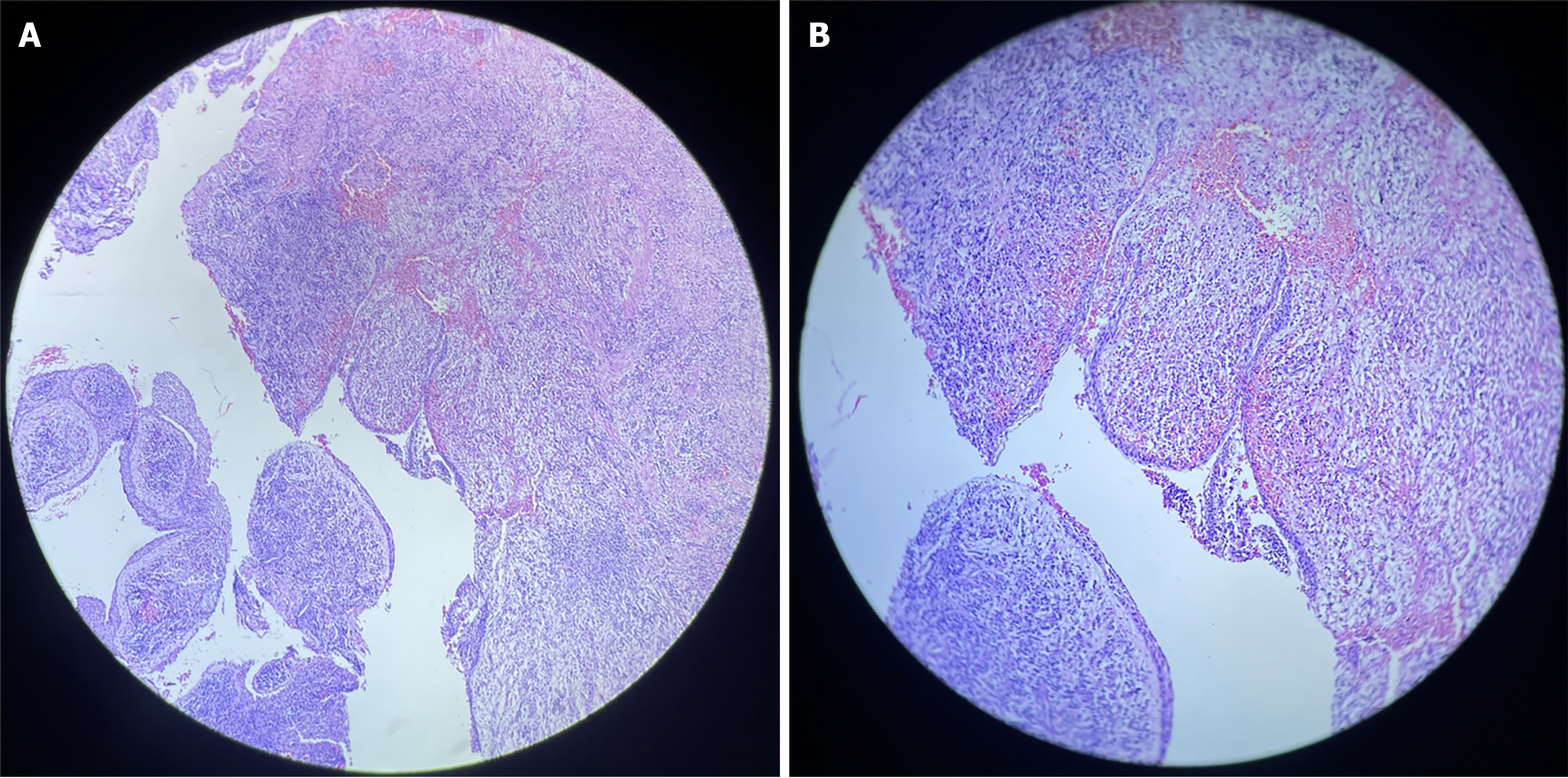
Figure 5 Histological examination.
A: Hematoxylin and eosin-stained histological sections exhibiting cavity lined by non-keratinized odontogenic epithelium with intracapsular projections beneath [Histological examination (H&E); × 20]; B: Photomicrograph showing intense, mixed inflammatory infiltrates and foamy macrophages in the fibrous capsule (H&E; × 40).
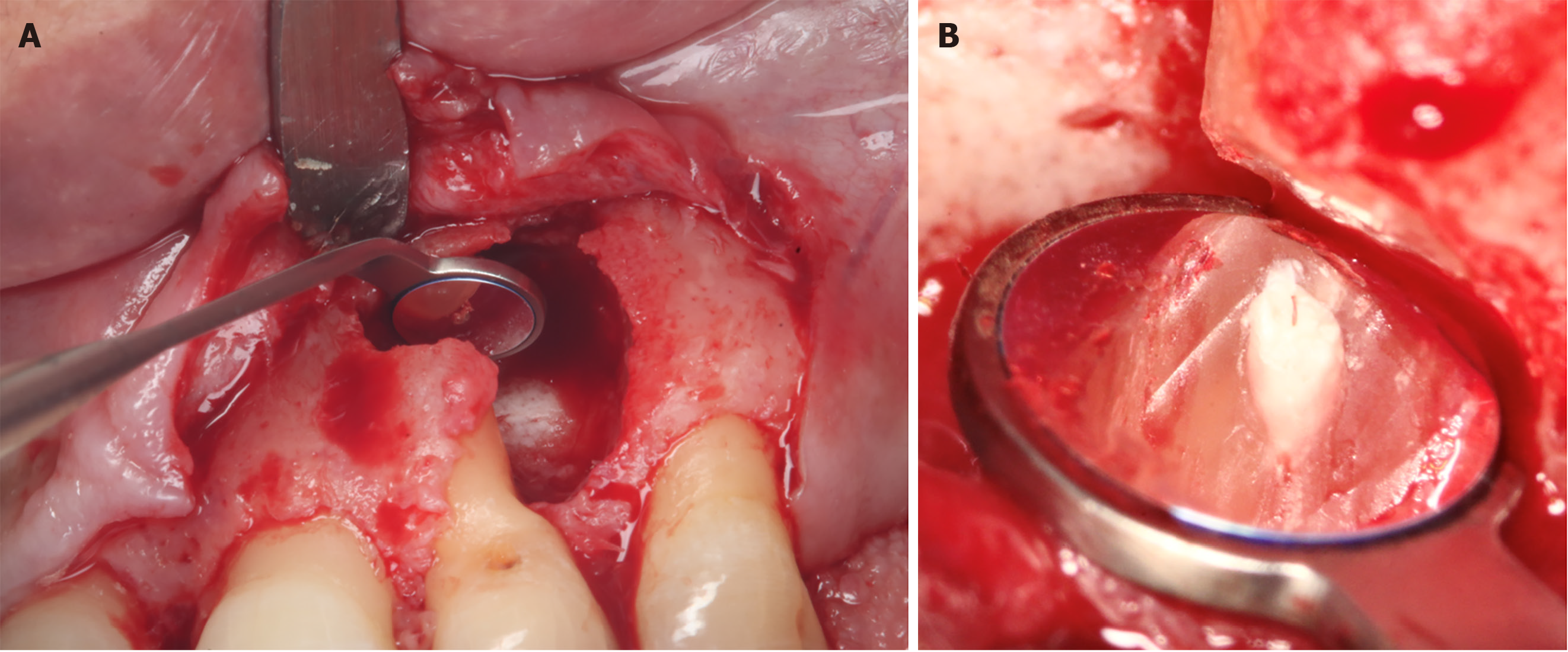
Figure 6 Intraoperative photography.
A: Complete enucleation of the lesion; B: Adequate sealing of the canal with biodentine, after apicectomy.
Figure 7 Immediate postoperative periapical radiograph.
Figure 8 Postoperative clinical view.
A: Immediate; B: After 10 d; C: After 4 months.
Figure 9 6 months postoperative cone-beam computed tomography view.
A: Sagittal; B: Axial; C: Coronal.
Figure 10 Six months postoperative three-dimensional model reconstruction.
- Citation: Gómez Mireles JC, Martínez Carrillo EK, Alcalá Barbosa K, Gutiérrez Cortés E, González Ramos J, González Gómez LA, Bayardo González RA, Lomelí Martínez SM. Microsurgical management of radicular cyst using guided tissue regeneration technique: A case report. World J Clin Cases 2024; 12(7): 1346-1355
- URL: https://www.wjgnet.com/2307-8960/full/v12/i7/1346.htm
- DOI: https://dx.doi.org/10.12998/wjcc.v12.i7.1346


