Copyright
©The Author(s) 2024.
World J Clin Cases. Mar 6, 2024; 12(7): 1305-1312
Published online Mar 6, 2024. doi: 10.12998/wjcc.v12.i7.1305
Published online Mar 6, 2024. doi: 10.12998/wjcc.v12.i7.1305
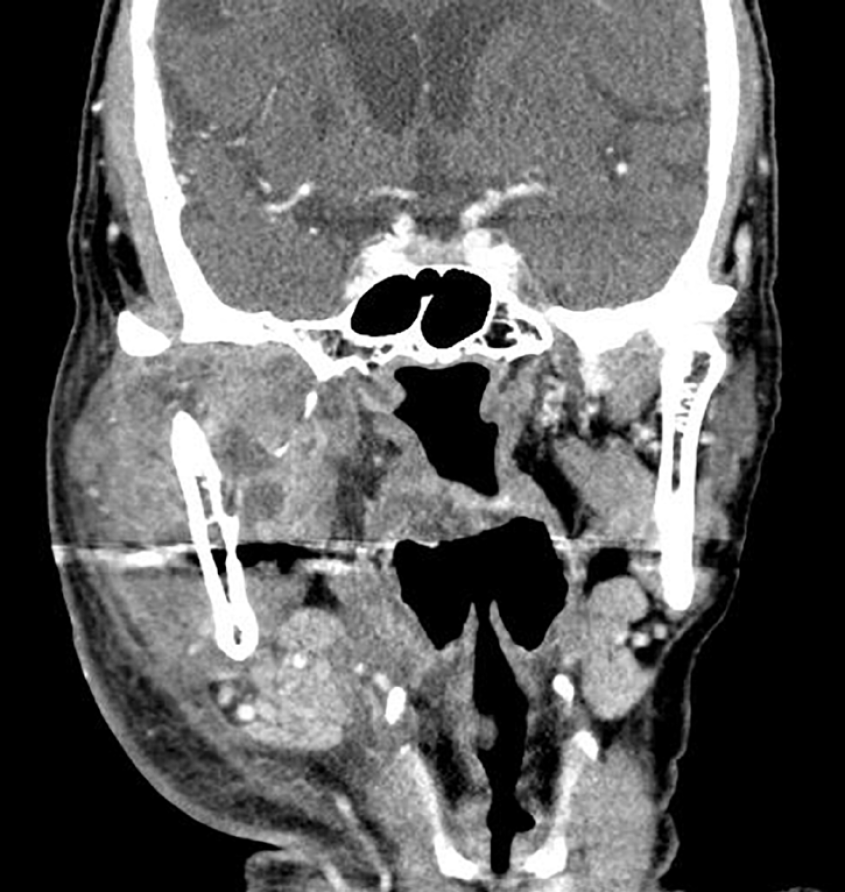
Figure 1 The contrast-enhanced computed tomography scan of the neck for Case 1.
The scan demonstrates diffuse thickening of the platysma muscle and subcutaneous fat stranding, along with significant swelling in the right mastication space and parotid space, indicative of cervical necrotizing fasciitis.
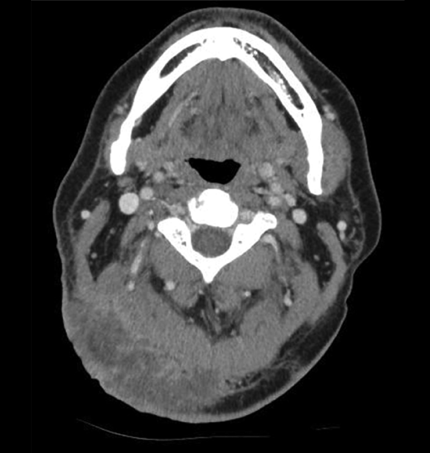
Figure 2 Contrast-enhanced computed tomography scan of the neck for Case 2.
The scan illustrated heterogeneous enhancement in the right occipital area and the adjacent posterior neck region. It also reveals diffuse thickening of the surrounding subcutaneous tissue and fat stranding all of which are consistent with the presence of cervical necrotizing fasciitis.
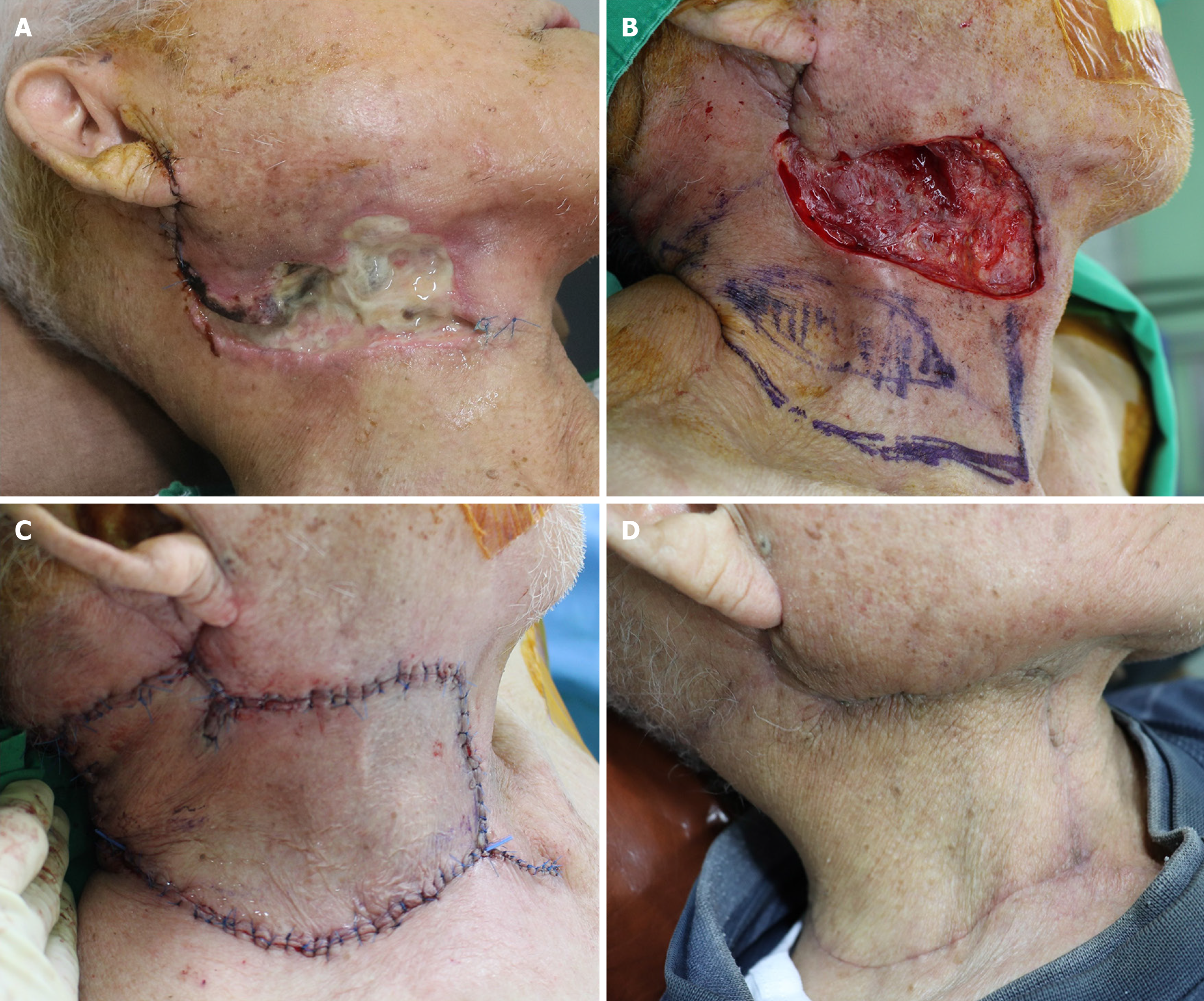
Figure 3 Clinical photographs of Case 1.
A: Skin necrosis caused by cervical necrotizing fasciitis in the right anterior neck area; B: The final post-debridement defect measuring 5.5 cm × 12 cm, along with the design of a keystone flap (KF) sized 8 cm × 19 cm, based on perforators from the superior thyroid artery; C: Successful coverage of the defect with the modified type II KF; D: Postoperative photographs taken after a 3-month follow-up.
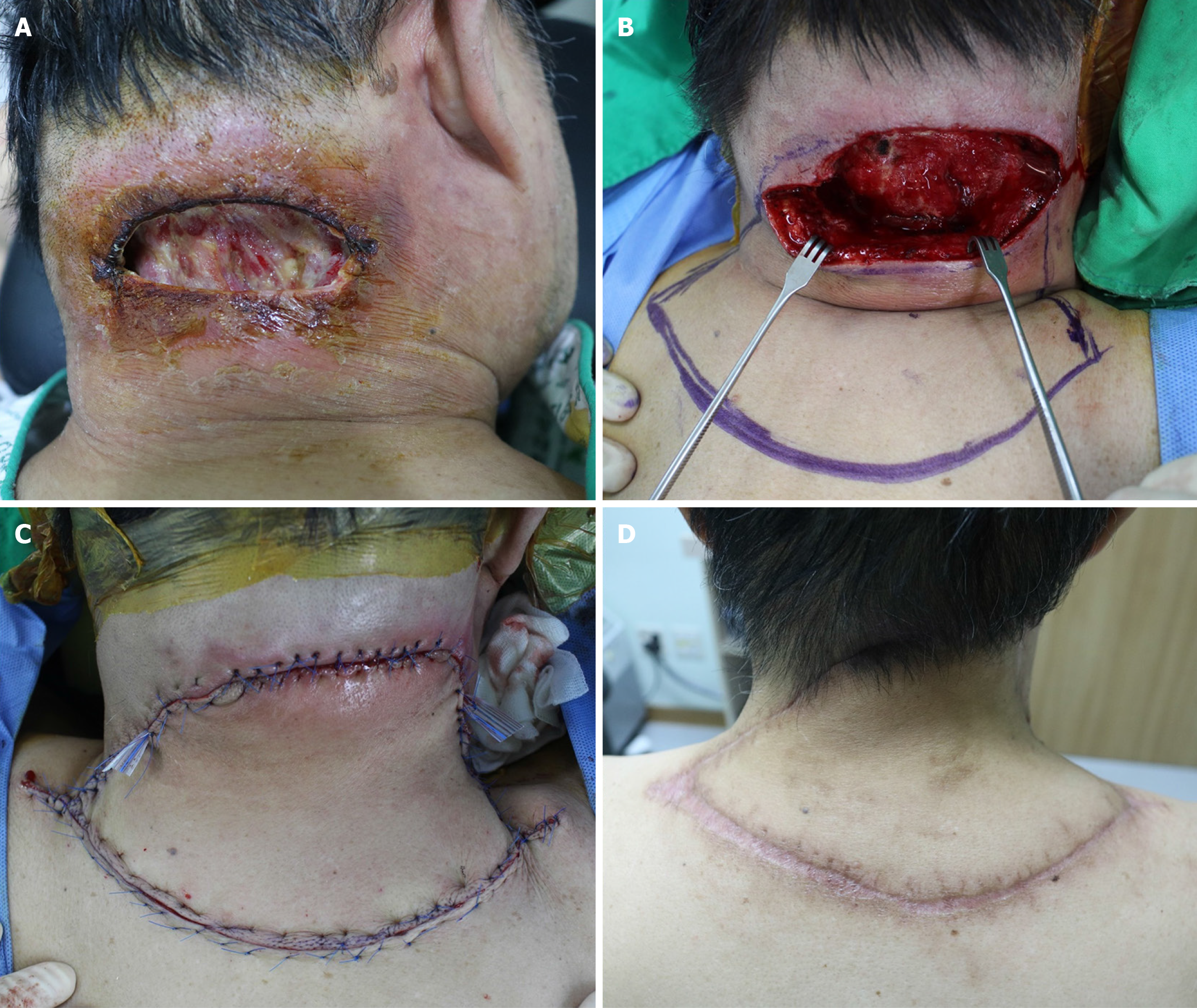
Figure 4 Clinical photographs of Case 2[8].
A: Skin necrosis resulting from cervical necrotizing fasciitis in the posterior neck area; B: The final post-debridement defect measuring 6 cm × 11 cm, alongside the design of a keystone flap (KF) sized 9 cm × 18 cm, based on perforators from the transverse cervical artery; C: Successful coverage of the defect with the modified Type II KF; D: Postoperative photographs taken after a 6-month follow-up. Citation: Kong YT, Kim J, Shin HW, Kim KN. Keystone flaps for coverage of defects in the posterior neck and lower occipital scalp: a retrospective clinical study. J Craniofac Surg 2021; 32: 1813-1816. Copyright ©The Authors 2021. Published by Wolters Kluwer Health, Inc.
Figure 5 Follow-up clinical photographs of Case 1.
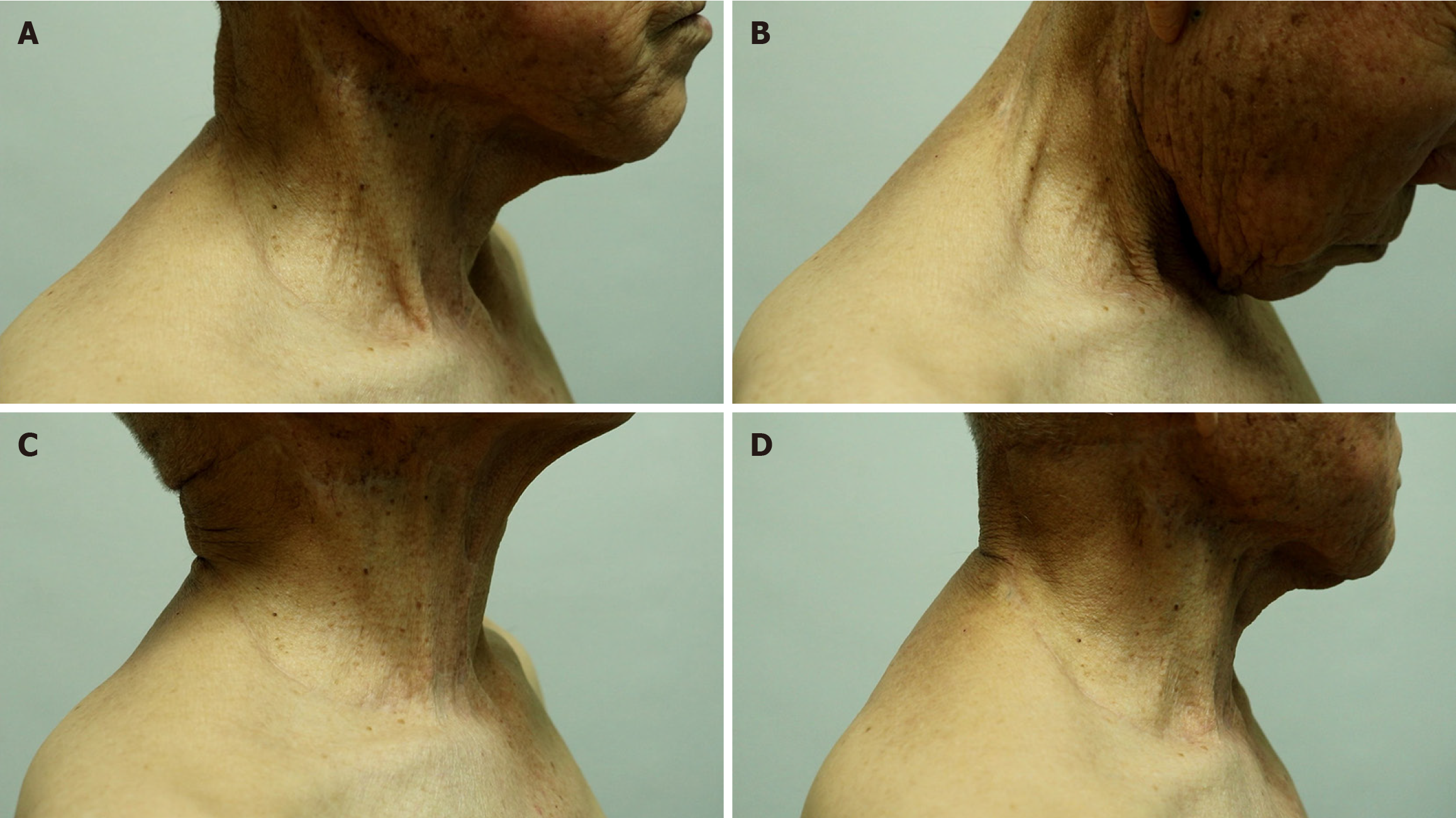
A-D: At the 7-month follow-up, no limitation of neck movement was observed.
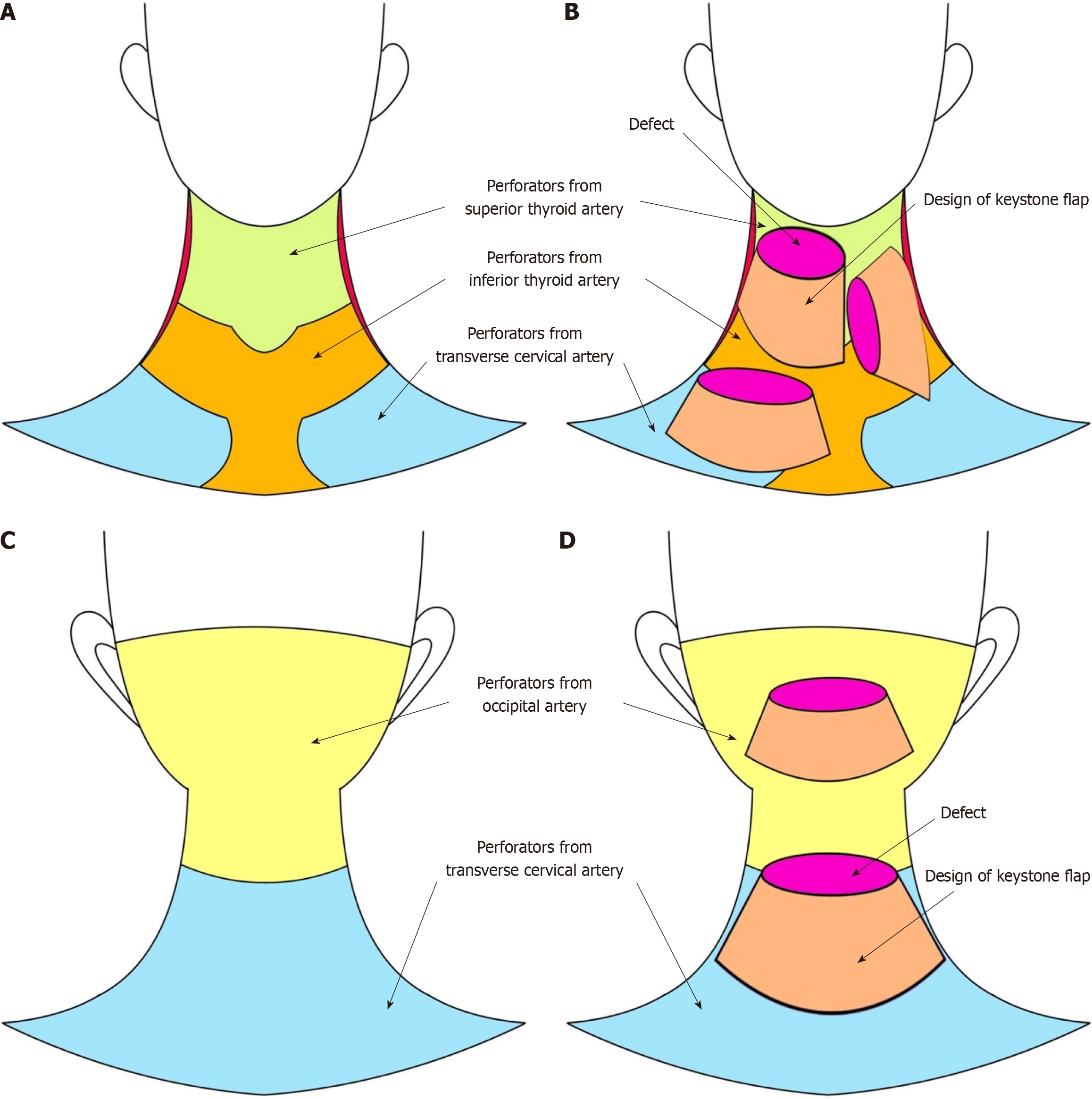
Figure 6 Illustrations of the regions of perforators in the neck area and the representative design of keystone flaps for neck defects.
A: Perforator territories of the anterior neck area. The chartreuse-colored territory represents the perforator-area from the superior thyroid artery, the mustard-colored territory represents the perforator-area from the inferior thyroid artery, and the sky-blue-colored territory represent the perforator-area from the transverse cervical artery; B: Representative designs of keystone flaps (KFs) for anterior neck defects. The magenta-colored ellipses represent defects, and the apricot-colored figures represent the design of KFs; C: Perforator territories of the posterior neck area. The lemon-colored territory represents the perforator-area from the occipital artery and the sky-blue-colored territory represent the perforator-area from the transverse cervical artery; D: Representative designs of KFs for posterior neck defects. The magenta-colored ellipses represent defects and the apricot-colored figures represent the design of KFs.
- Citation: Cho W, Jang EA, Kim KN. Reconstruction of cervical necrotizing fasciitis defect with the modified keystone flap technique: Two case reports. World J Clin Cases 2024; 12(7): 1305-1312
- URL: https://www.wjgnet.com/2307-8960/full/v12/i7/1305.htm
- DOI: https://dx.doi.org/10.12998/wjcc.v12.i7.1305


