Copyright
©The Author(s) 2024.
World J Clin Cases. Mar 6, 2024; 12(7): 1296-1304
Published online Mar 6, 2024. doi: 10.12998/wjcc.v12.i7.1296
Published online Mar 6, 2024. doi: 10.12998/wjcc.v12.i7.1296
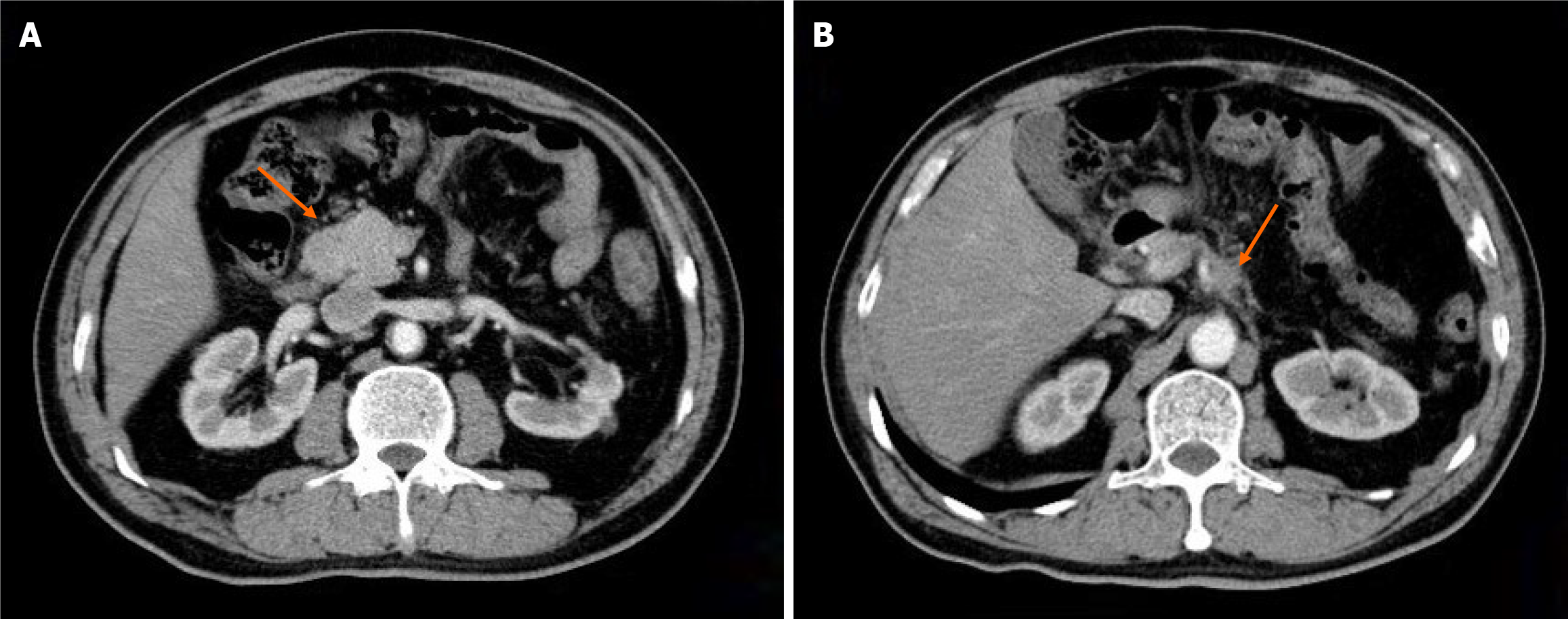
Figure 1 Computed tomography.
A: Computed tomography revealed one reinforcing nodule anterior to the descending portion of the duodenum; B: Considered metastatic; soft tissue shadows adjacent to the celiac trunk.
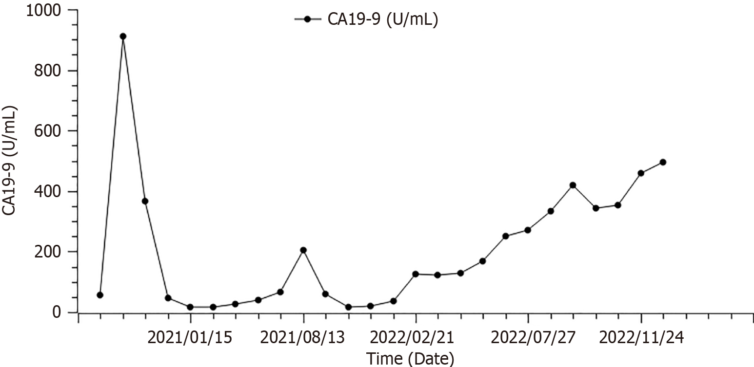
Figure 2 Change in carbohydrate antigen 19-9 over time (case 1).
CA19-9: Carbohydrate antigen 19-9.
Figure 3 Computed tomography scan of patient 1 after taking fruquintinib remained a stable disease for 10 months.
A: Computed tomography (CT) scan of the target lesion, February 2022; B: CT scan of the target lesion, April 2022; C: CT scan of the target lesion, August 2022; D: CT scan of the target lesion, December 2022.
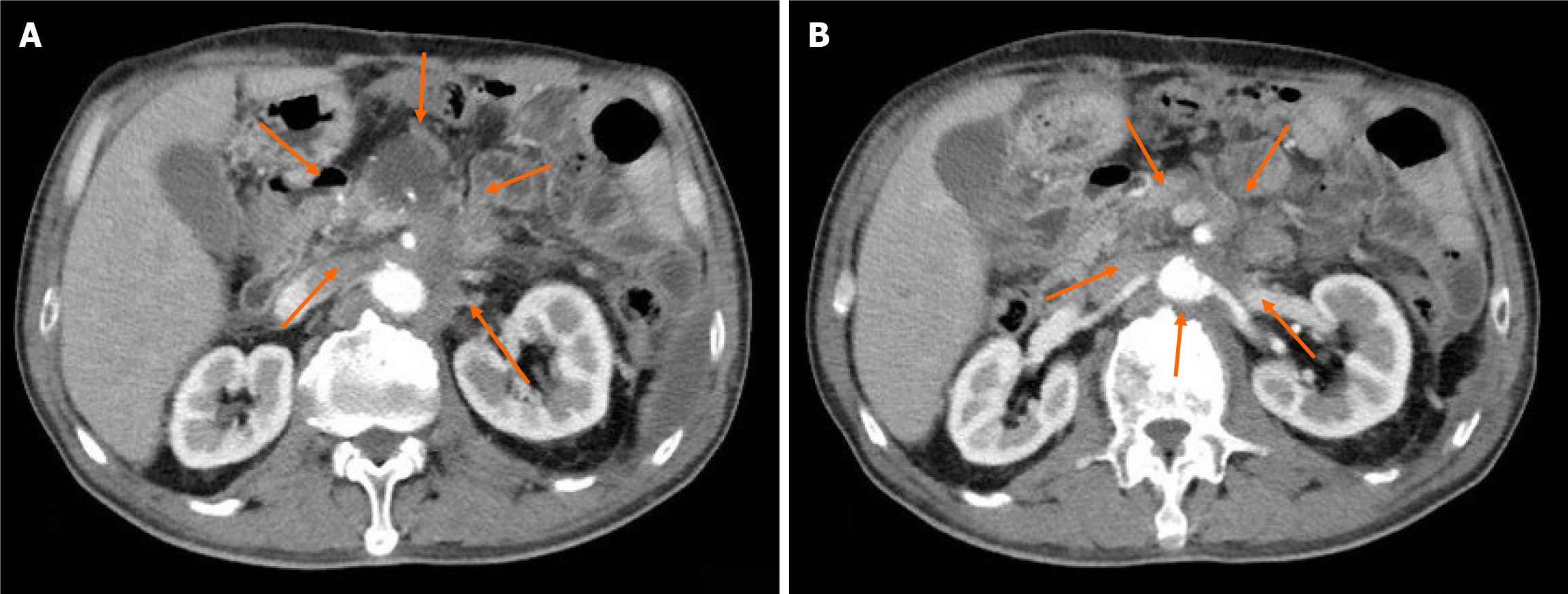
Figure 4 Computed tomography revealed postoperative changes in pancreatic cancer.
A: Revealing cystic foci near the portal vein, multiple abdominopelvic nodules; B: Potential metastasis, and enlarged abdominal lymph nodes.
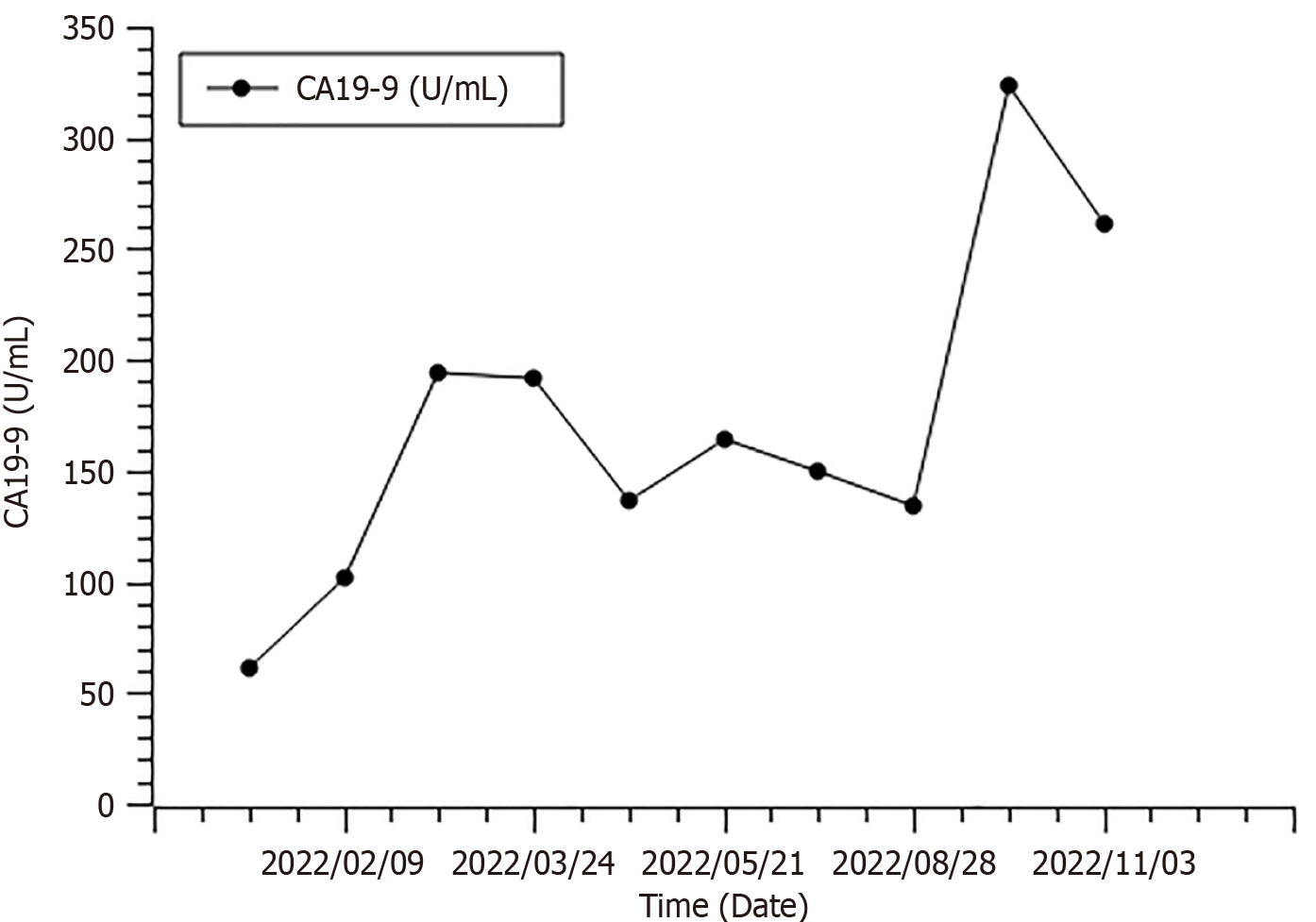
Figure 5 Change in carbohydrate antigen 19-9 over time (case 2).
CA19-9: Carbohydrate antigen 19-9.
Figure 6 Computed tomography scan of case 2.
The computed tomography (CT) scan revealed a continuous partial response from February 23, 2022, to December 7, 2022. A: CT scan showed the target lesion of 51 mm × 46 mm, February 2022; B: CT scan showed the target lesion of 34 mm × 38 mm, April 2022; C: CT scan showed the target lesion of 33 mm × 38 mm, June 2022; D: CT scan showed the target lesion of 35 mm × 31 mm, October 2022; E: CT scan showed the target lesion of 36 mm × 31 mm, December 2022.
- Citation: Wu D, Wang Q, Yan S, Sun X, Qin Y, Yuan M, Wang NY, Huang XT. Extended survival with metastatic pancreatic cancer under fruquintinib treatment after failed chemotherapy: Two case reports. World J Clin Cases 2024; 12(7): 1296-1304
- URL: https://www.wjgnet.com/2307-8960/full/v12/i7/1296.htm
- DOI: https://dx.doi.org/10.12998/wjcc.v12.i7.1296


