Copyright
©The Author(s) 2024.
World J Clin Cases. Feb 26, 2024; 12(6): 1182-1189
Published online Feb 26, 2024. doi: 10.12998/wjcc.v12.i6.1182
Published online Feb 26, 2024. doi: 10.12998/wjcc.v12.i6.1182
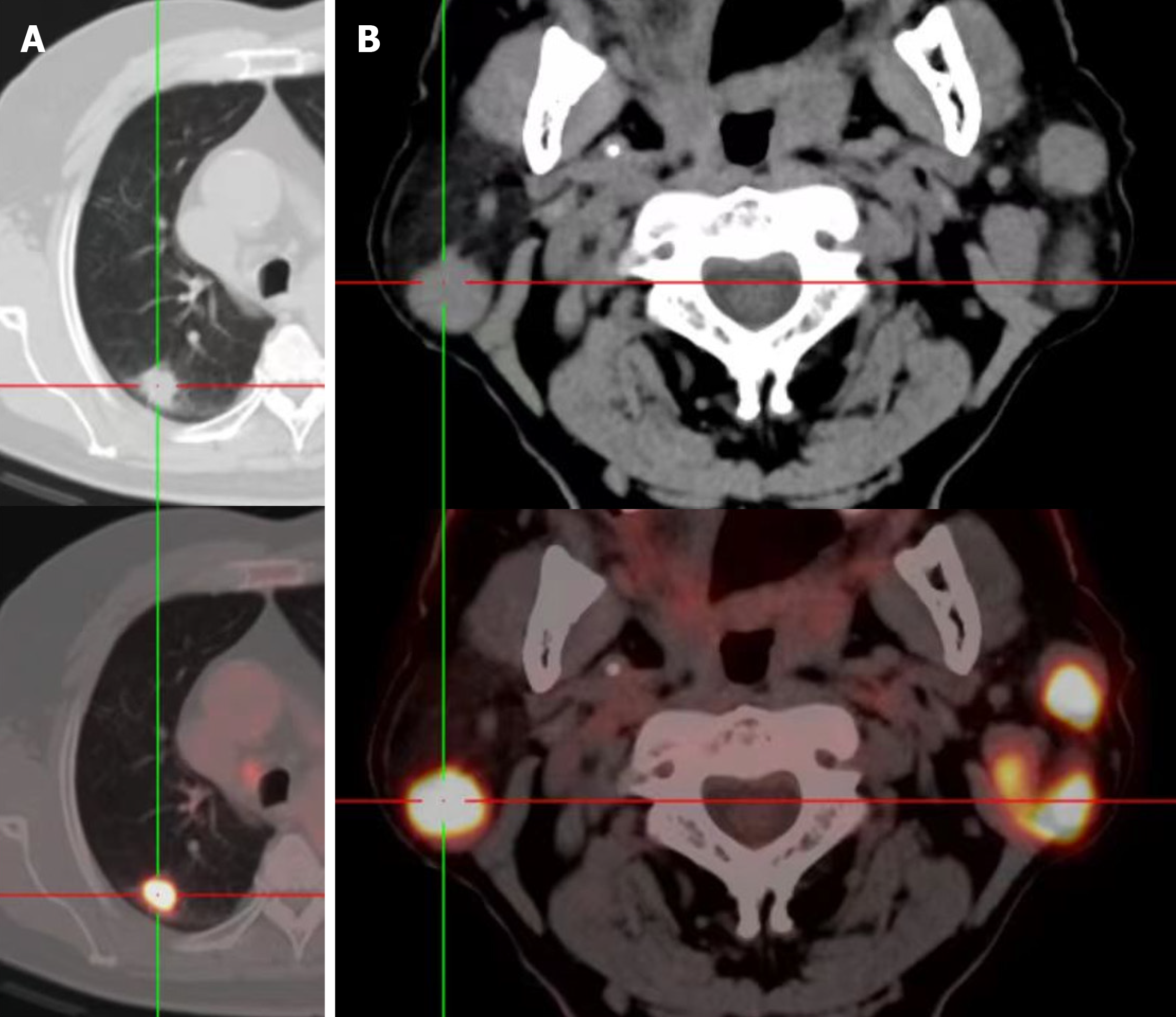
Figure 1 18F fluorodeoxyglucose values shown by positron emission tomography.
A: Superior lobe of right lung (18.2); B: Bilateral parotid gland (27.3).
Figure 2 Histopathological examination.
A: Lung (× 400); B: Parotid gland (× 400).
- Citation: Yan RX, Dou LB, Wang ZJ, Qiao X, Ji HH, Zhang YC. Parotid metastasis of rare lung adenocarcinoma: A case report. World J Clin Cases 2024; 12(6): 1182-1189
- URL: https://www.wjgnet.com/2307-8960/full/v12/i6/1182.htm
- DOI: https://dx.doi.org/10.12998/wjcc.v12.i6.1182


