Copyright
©The Author(s) 2024.
World J Clin Cases. Feb 26, 2024; 12(6): 1138-1143
Published online Feb 26, 2024. doi: 10.12998/wjcc.v12.i6.1138
Published online Feb 26, 2024. doi: 10.12998/wjcc.v12.i6.1138
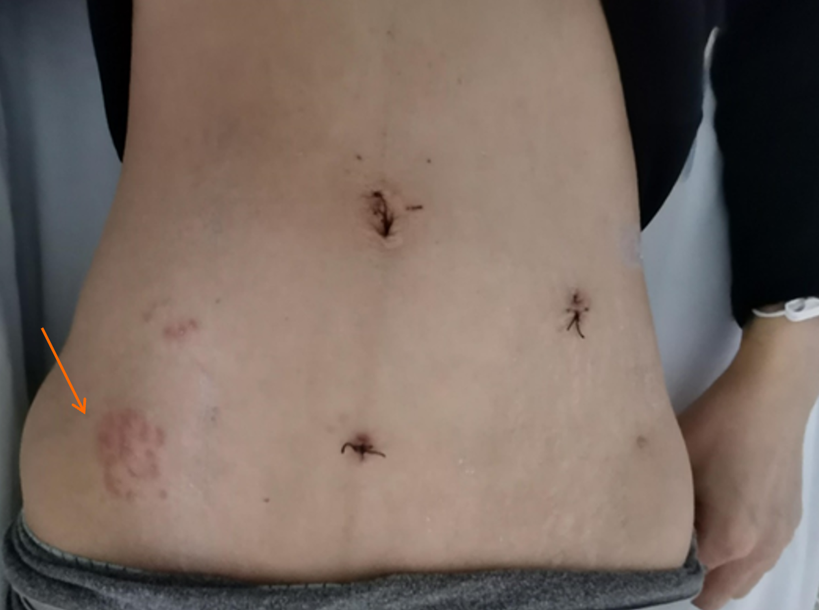
Figure 1 Clusters of red rashes.
Clusters of red rashes can be seen on the patient's right lower abdomen (as shown by the arrow).
Figure 2 Abdominal upright plain film.
Gas accumulation in the abdominal intestine, and scattered fluid levels are seen, indicating intestinal obstruction.
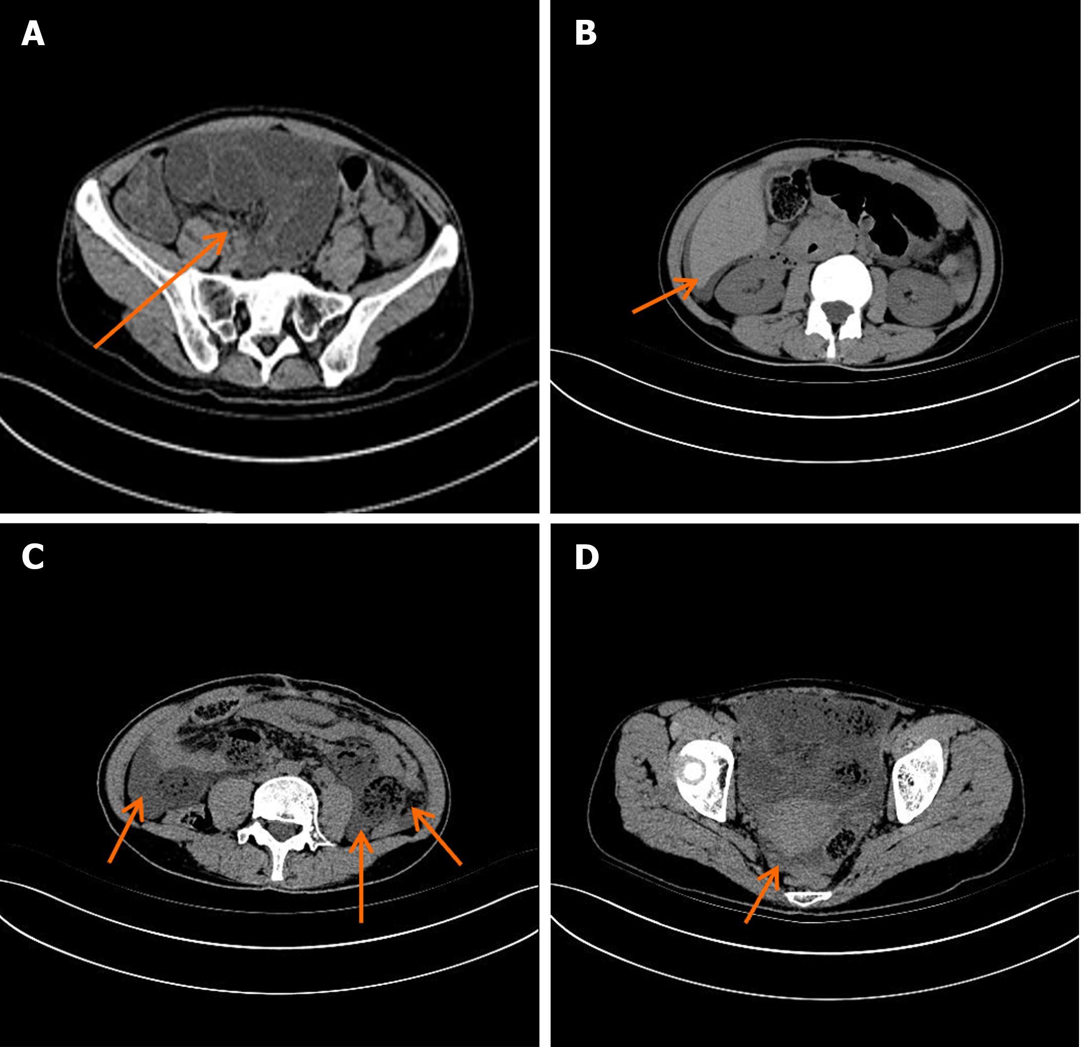
Figure 3 Representative whole-abdominal computed tomography images.
A: Significant dilation and fluid accumulation of the bowel; B: An arc-shaped hypodense shadow under the capsule of the liver at the point indicated by the arrow; C: Fluid accumulation in abdominal cavity by the arrow; D: Fluid accumulation in pelvic cavity by the arrow.
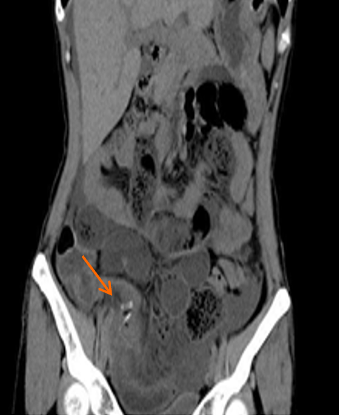
Figure 4 Representative coronal plane whole-abdominal computed tomography image.
The position of the Hem-Lock clamp during the patient's previous laparoscopic appendectomy.
- Citation: Dong ZY, Shi RX, Song XB, Du MY, Wang JJ. Postoperative abdominal herpes zoster complicated by intestinal obstruction: A case report. World J Clin Cases 2024; 12(6): 1138-1143
- URL: https://www.wjgnet.com/2307-8960/full/v12/i6/1138.htm
- DOI: https://dx.doi.org/10.12998/wjcc.v12.i6.1138


