Copyright
©The Author(s) 2024.
World J Clin Cases. Feb 26, 2024; 12(6): 1130-1137
Published online Feb 26, 2024. doi: 10.12998/wjcc.v12.i6.1130
Published online Feb 26, 2024. doi: 10.12998/wjcc.v12.i6.1130
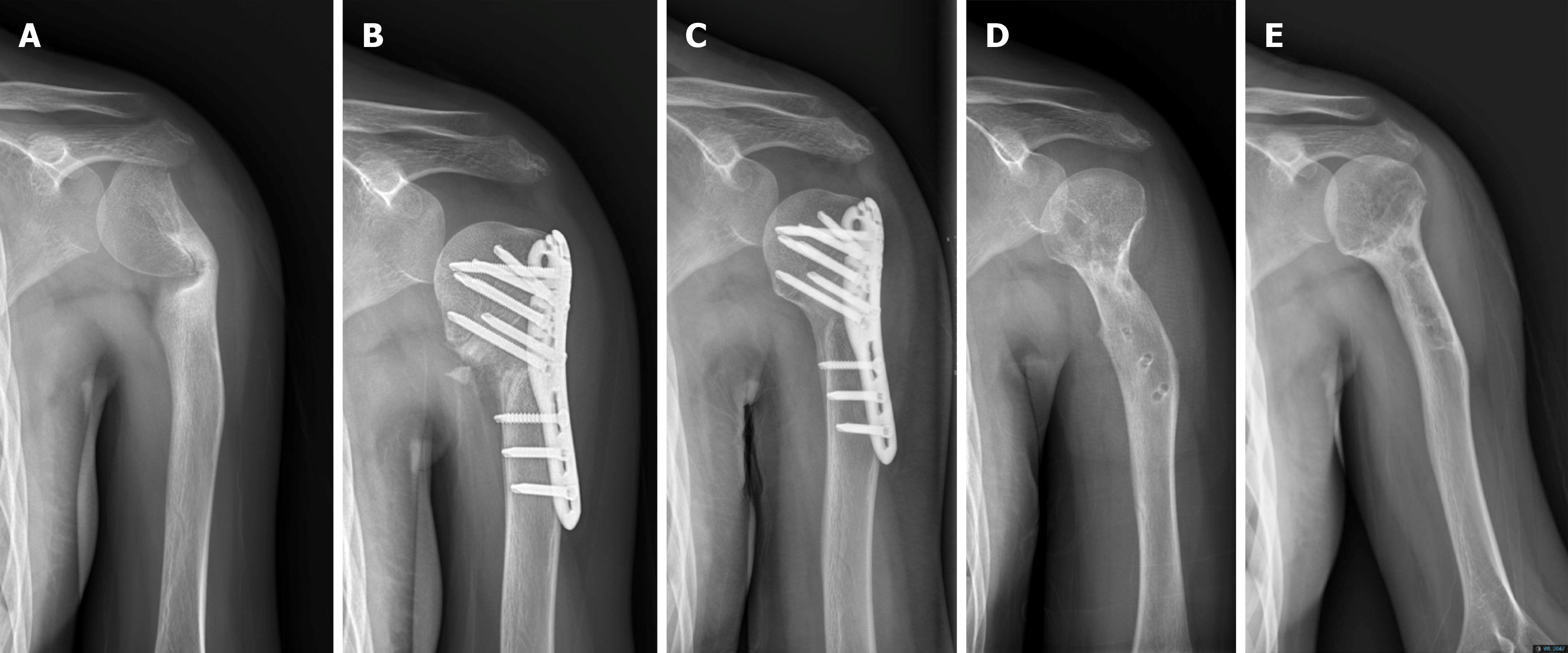
Figure 1 Series anteroposterior plain radiographs of the left humerus.
A: Preoperative anteroposterior plain radiographs of the left humerus at initial presentation; B: 1-month postoperative anteroposterior plain radiographs of the left humerus; C: 1-year post-operative anteroposterior plain radiographs of the left humerus; D: Anteroposterior plain radiographs of the left humerus at 1-year post-removal of internal fixator; E: 1-year post-removal of internal fixator surgery.
Figure 2 Preoperative magnetic resonance images and pathological results of the patient.
Preoperative T2-weighted magnetic resonance images of the left humerus showed a marked deformity of the humeral head and a potential malunion of the displaced fracture in the left humeral neck.
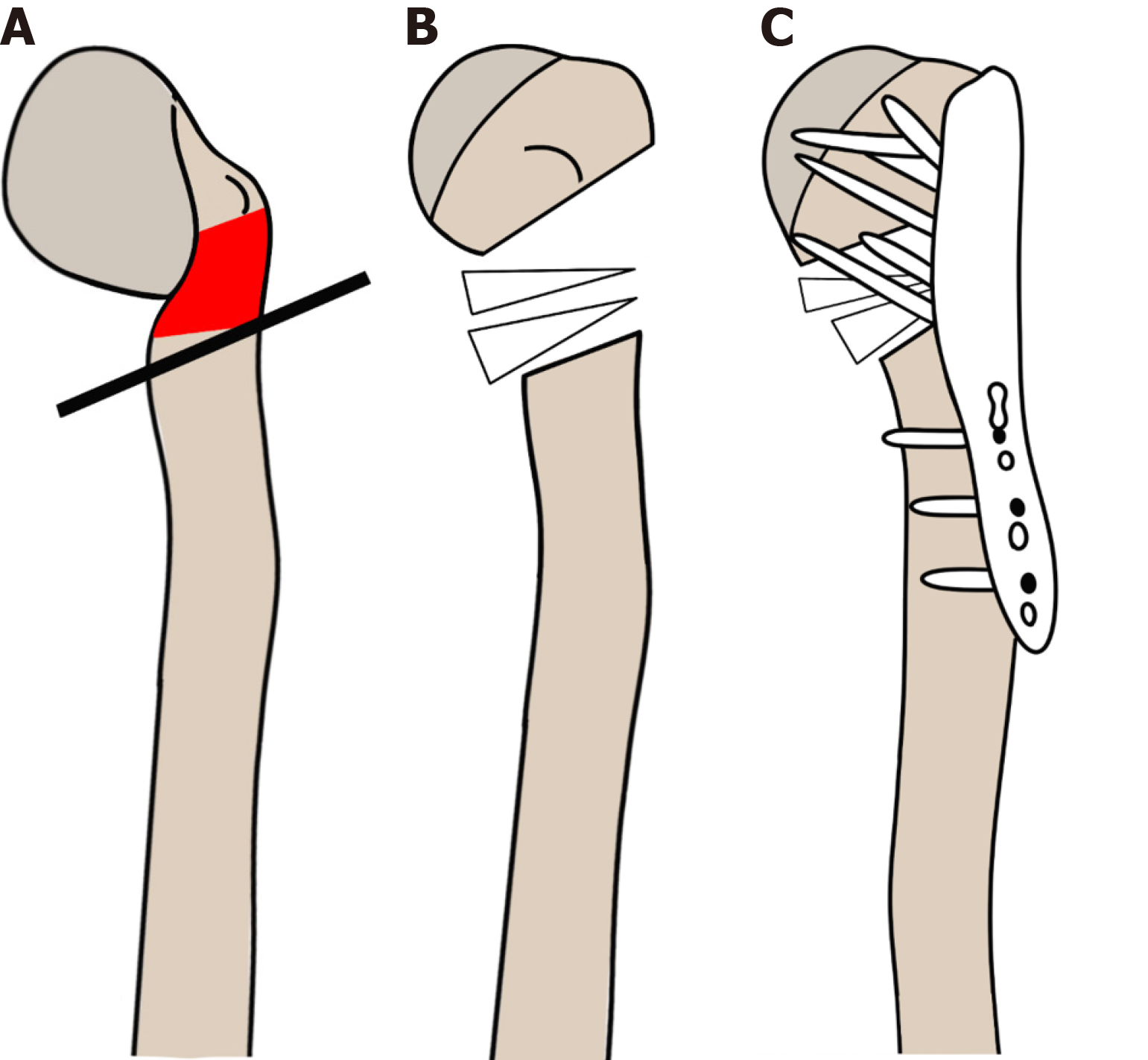
Figure 3 Surgical procedure for acute correction of the proximal humerus for the varus deformity.
The red area indicates the location of the simple bone cyst (SBC). A: Osteotomy was performed in the most angulated area, removing the deformity site of the SBC; B: The gap was filled with allograft (donor femoral head); C: The proximal humerus was fixed with an anatomical locking plate (Depuy-Synthesis®, Raynham, MA, United States) in a valgus position.
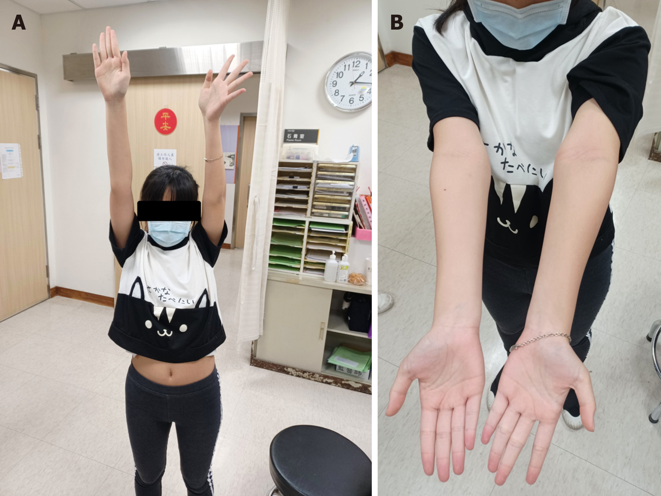
Figure 4 Two-week postoperative photos of the patient.
A: Full active range of motion of the left shoulder; B: About 2-3 cm length discrepancy on the affected side.
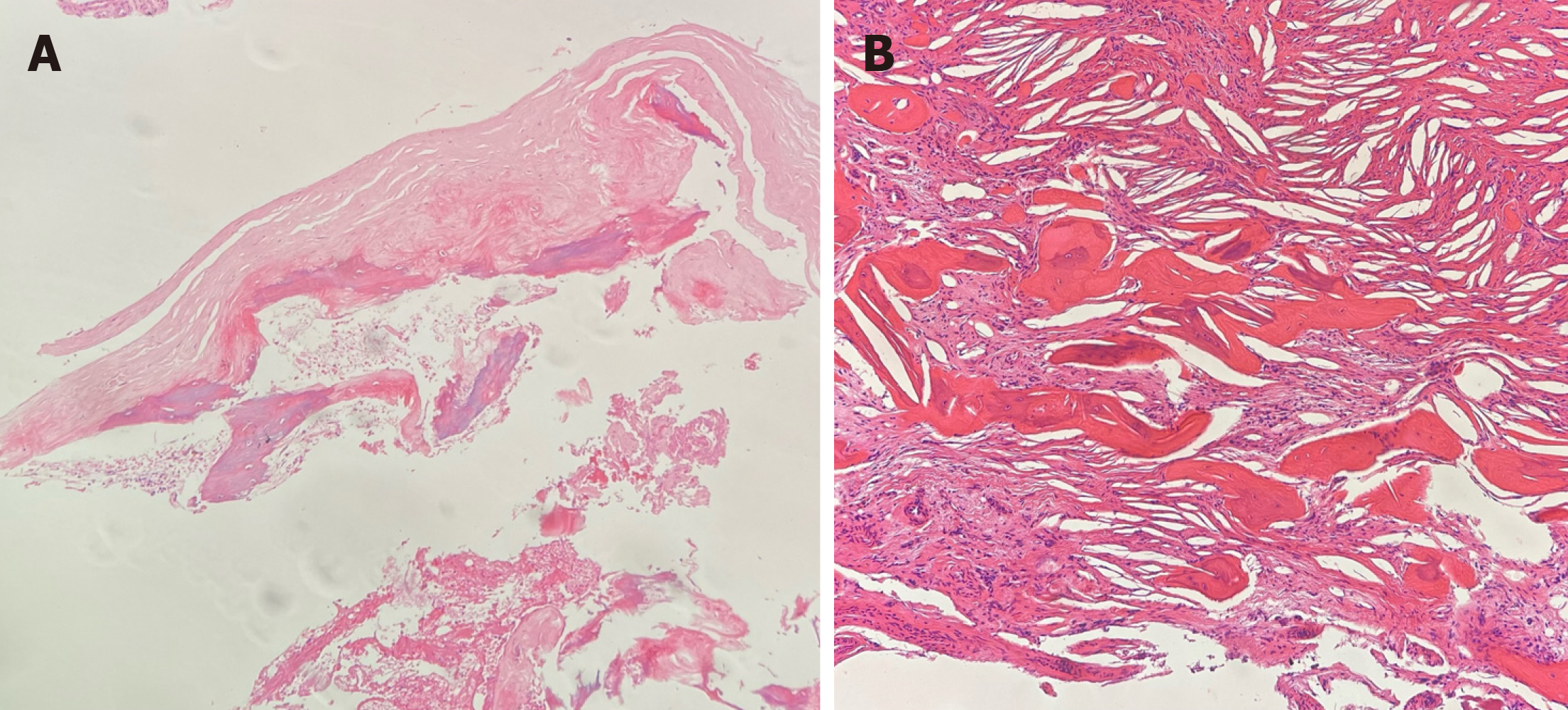
Figure 5 Pathological results of the patient.
A: Histological results showed fibrous cyst wall with fibrin-like material and calcification; B: Histological results showed cystic wall with cholesterol clefts and surrounding some new bone formation, all of which may be indicative of a diagnosis of a simple bone cyst.
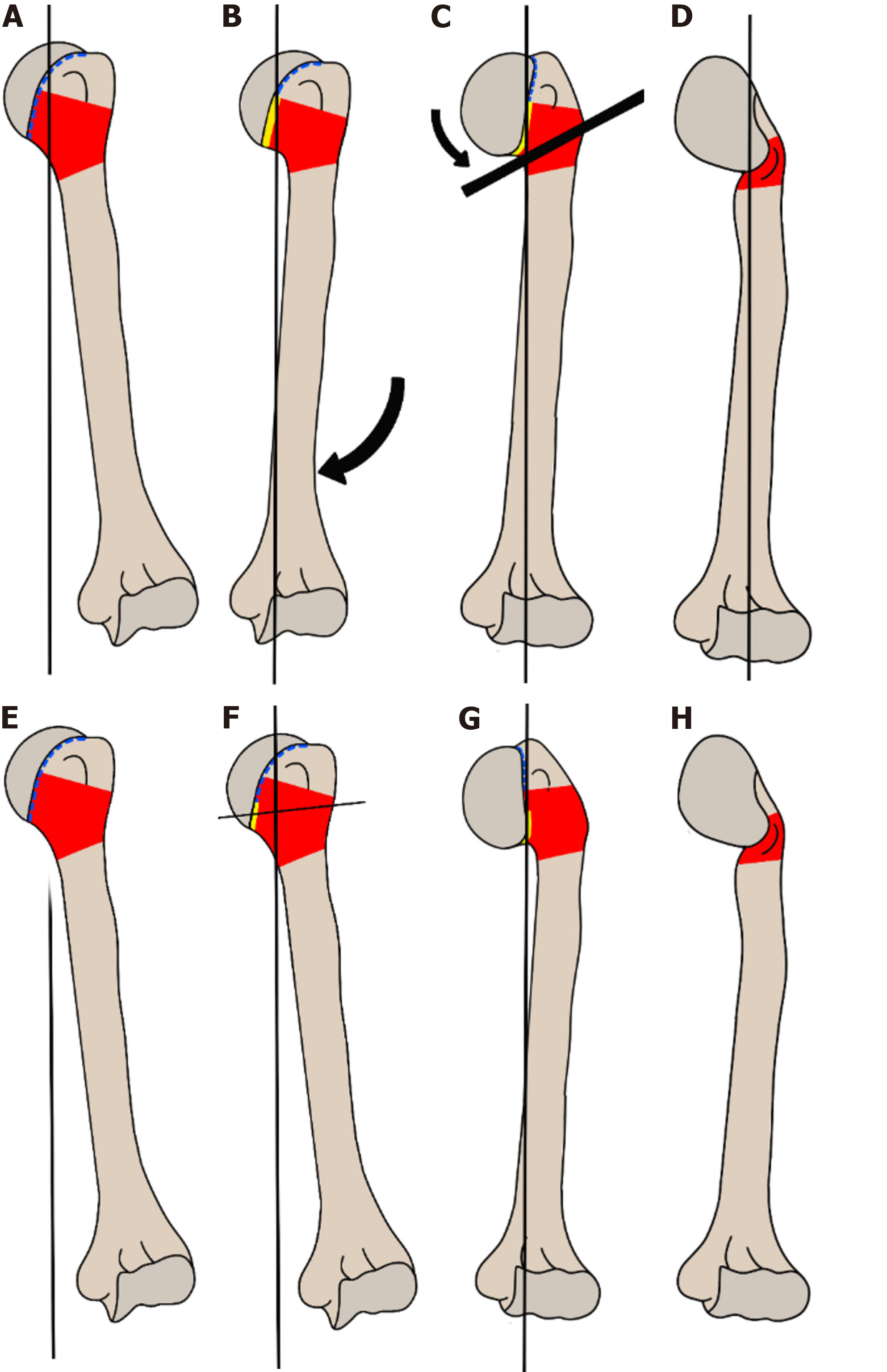
Figure 6 Two hypotheses for the etiology of our patient with simple bone cyst who presented with a pathological fracture, angular deformity, and limb length discrepancy.
The red area represents the location of simple bone cyst (SBC); the blue dotted line represents the epiphysis; and the yellow line indicates the affected epiphysis. A–D: The first hypothesis. A: The proximal humeral simple bone cyst extends through the physis on the medial side; B: The SBC subsequently caused growth arrest and varus deformity of the humerus; C: Due to a proximal humerus fracture, additional varus angulation of the humeral head occurs; D: SBC presenting with a pathological fracture, complicated with malunion of the displaced fracture, varus deformity and limb length discrepancy. E–H: The second hypothesis. E: A proximal humeral simple bone cyst develops; F: Repetitive pathological fractures due to SBC damaged the medial epiphysis of the proximal humerus and varus humeral head angulation was established as a consequence; G: The humerus develops varus deformity and a length discrepancy due to a damaged epiphysis; H: SBC presenting with a pathological fracture, complicated with malunion of the displaced fracture, varus deformity and limb length discrepancy.
- Citation: Lin CS, Lin SM, Rwei SP, Chen CW, Lan TY. Simple bone cysts of the proximal humerus presented with limb length discrepancy: A case report. World J Clin Cases 2024; 12(6): 1130-1137
- URL: https://www.wjgnet.com/2307-8960/full/v12/i6/1130.htm
- DOI: https://dx.doi.org/10.12998/wjcc.v12.i6.1130


