Copyright
©The Author(s) 2024.
World J Clin Cases. Feb 26, 2024; 12(6): 1045-1049
Published online Feb 26, 2024. doi: 10.12998/wjcc.v12.i6.1045
Published online Feb 26, 2024. doi: 10.12998/wjcc.v12.i6.1045
Figure 1 An irregular mass of tumor tissue in the axillary adipose tissue.
There is no lymphoid tissue at the periphery or within the tumor mass. This will qualify for designation of tumor deposits in axillary fat (H&E, × 40).
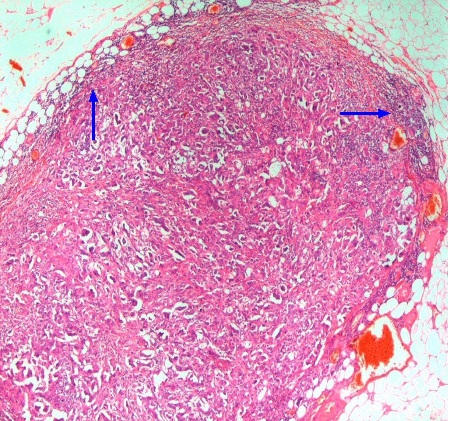
Figure 2 In this case, the tumor mass is exhibiting ovoid or reniform configuration consistent with lymph node metastasis.
Focally, some lymphoid tissue can be seen at the periphery of the tumor mass (arrows). This will not qualify for the designation of tumor deposits (H&E, × 40).
Figure 3 Another example of lymph node metastasis in the axilla.
A variably thick rim of lymphoid tissue can be seen at the periphery of the tumor mass (arrows). This will also not qualify for tumor deposits designation (H&E, ×40).
- Citation: Mubarak M, Rashid R, Shakeel S. Tumor deposits in axillary adipose tissue in patients with breast cancer: Do they matter? World J Clin Cases 2024; 12(6): 1045-1049
- URL: https://www.wjgnet.com/2307-8960/full/v12/i6/1045.htm
- DOI: https://dx.doi.org/10.12998/wjcc.v12.i6.1045


