Copyright
©The Author(s) 2024.
World J Clin Cases. Feb 16, 2024; 12(5): 995-1003
Published online Feb 16, 2024. doi: 10.12998/wjcc.v12.i5.995
Published online Feb 16, 2024. doi: 10.12998/wjcc.v12.i5.995
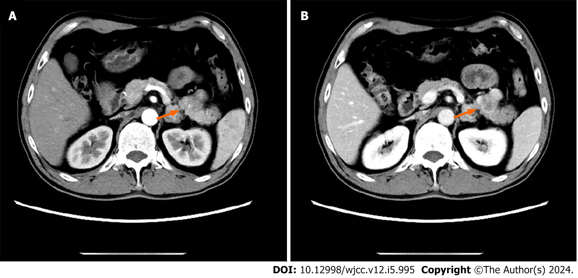
Figure 1 Abdominal computed tomography scan showing a 5.
52 cm × 2.82 cm × 2 cm mass in the pancreas (orange arrows). A: No enhancement in the arterial region. B: Heterogeneous enhancement in the venous area.
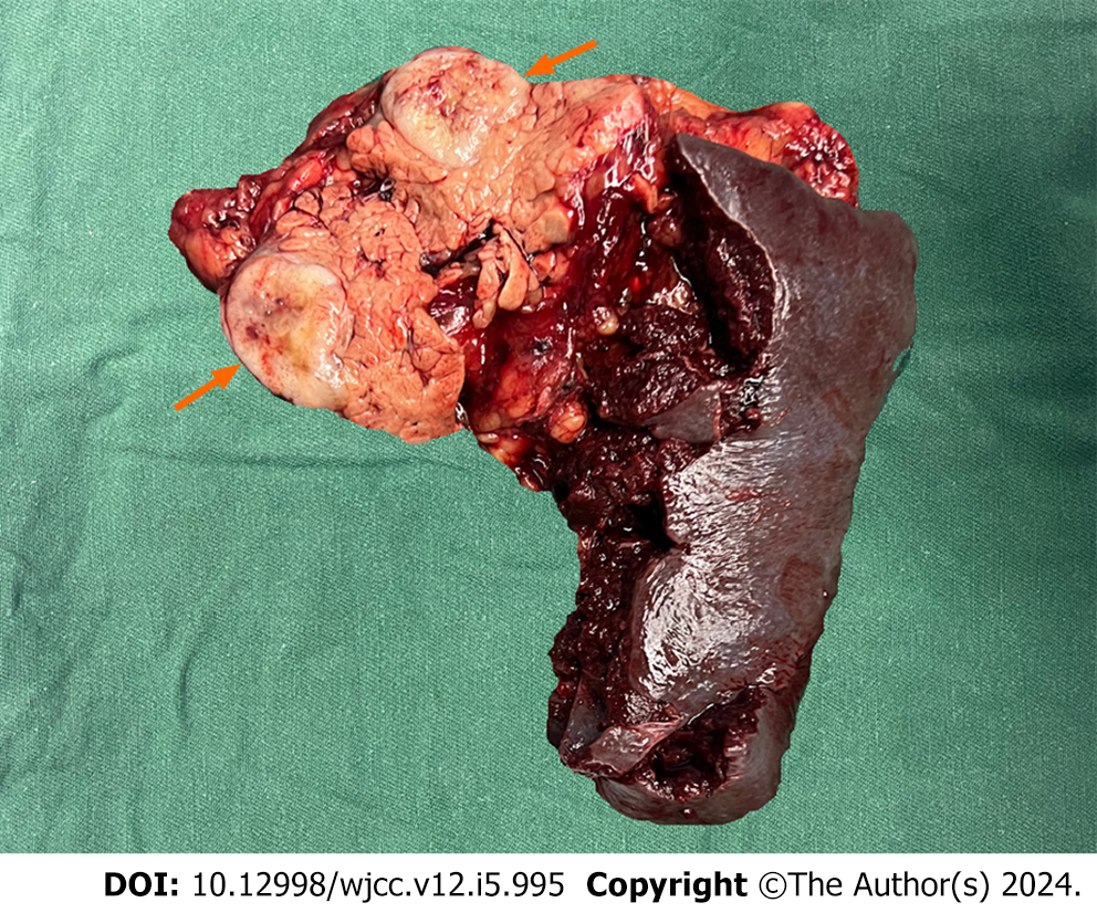
Figure 2 Postoperative surgical specimen: Pancreatic tail and spleen (tumor cut open chart) (orange arrows).
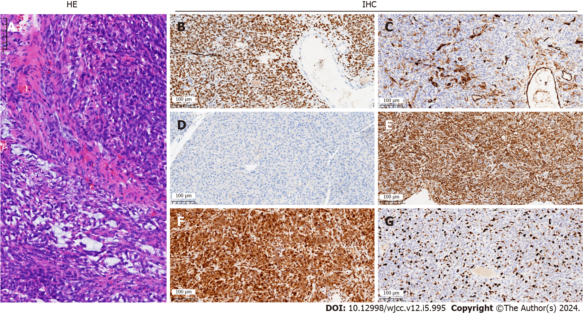
Figure 3 Representative results of hematoxylin and eosin and immunohistochemical staining of surgical specimens of solitary fibrous tumor of the pancreas.
A: Hematoxylin and Eosin staining (hematoxylin and Shuhong); B: Immunohistochemistry (original magnifcation of × 400) signal transducer and activator of transcription 6; C: CD34; D: CD99; E: Vimentin; F: Vimentin; G: Ki-67.
- Citation: Wang WW, Zhou SP, Wu X, Wang LL, Ruan Y, Lu J, Li HL, Ni XL, Qiu LL, Zhou XH. Imaging, pathology, and diagnosis of solitary fibrous tumor of the pancreas: A case report and review of literature. World J Clin Cases 2024; 12(5): 995-1003
- URL: https://www.wjgnet.com/2307-8960/full/v12/i5/995.htm
- DOI: https://dx.doi.org/10.12998/wjcc.v12.i5.995


