Copyright
©The Author(s) 2024.
World J Clin Cases. Feb 16, 2024; 12(5): 988-994
Published online Feb 16, 2024. doi: 10.12998/wjcc.v12.i5.988
Published online Feb 16, 2024. doi: 10.12998/wjcc.v12.i5.988
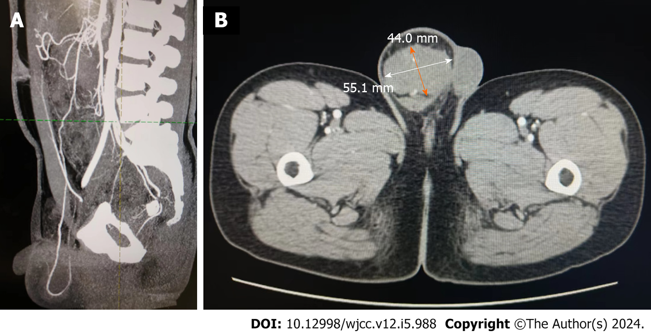
Figure 1 Computed tomography findings.
A: The blood supply of scrotal masses is from the omentum or mesenteric vessels in the abdominal cavity; B: The scrotal mass’s diameter is 44.0 mm × 55.1 mm.
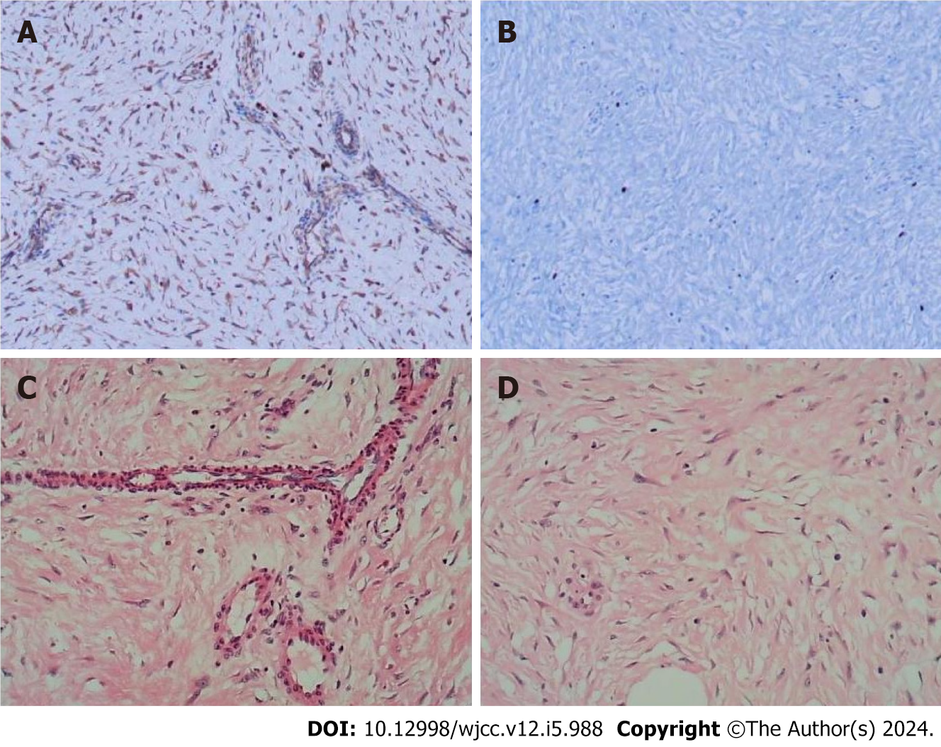
Figure 2 Pathological sections were stained.
A and B: Image shows immunohistochemical staining; C and D: Image shows hematoxylin and eosin staining.
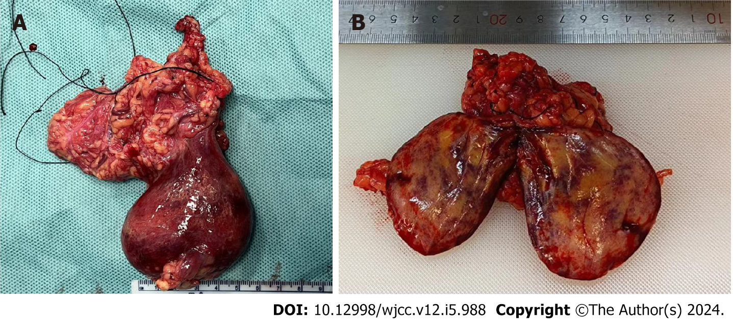
Figure 3 Resected scrotal mass: Originating from the greater omentum.
A: The mass envelope was complete, and the boundary between the mass and testis was clear; B: Cross-sectional view of the tumor: Slightly hard, gray-yellow, solid mass.
- Citation: Zhou P, Jin CH, Shi Y, Ma GQ, Wu WH, Wang Y, Cai K, Fan WF, Wang TB. Omental fibroma combined with right indirect inguinal hernia masquerades as a scrotal tumor: A case report. World J Clin Cases 2024; 12(5): 988-994
- URL: https://www.wjgnet.com/2307-8960/full/v12/i5/988.htm
- DOI: https://dx.doi.org/10.12998/wjcc.v12.i5.988


