Copyright
©The Author(s) 2024.
World J Clin Cases. Feb 16, 2024; 12(5): 973-979
Published online Feb 16, 2024. doi: 10.12998/wjcc.v12.i5.973
Published online Feb 16, 2024. doi: 10.12998/wjcc.v12.i5.973
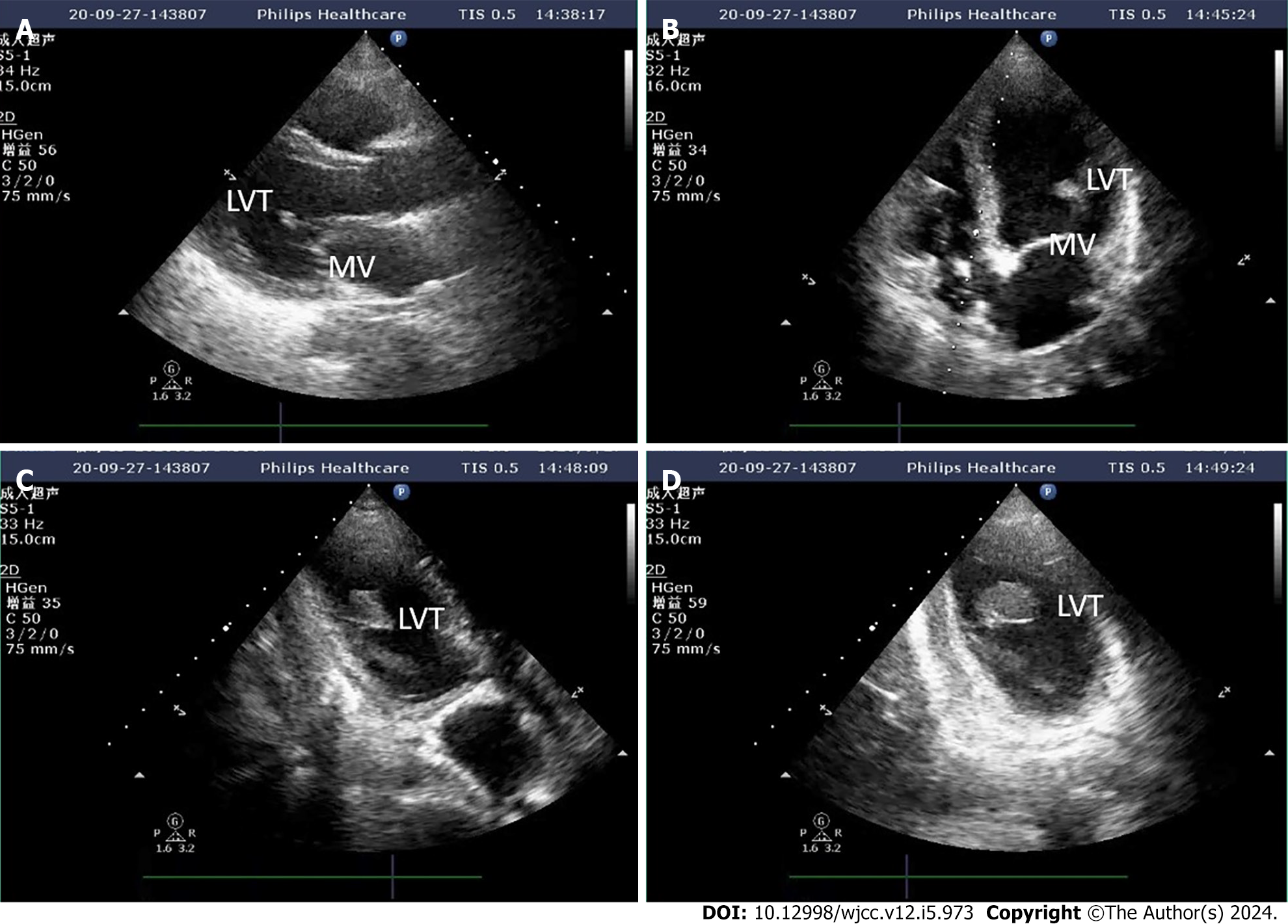
Figure 1 Left ventricular thrombosis indicated by transthoracic ultrasonography.
A: The long axis of the left ventricle (LV) beside the sternum indicates suspected thrombosis attached to the mitral valve; B: Apical four-chamber heart suggests suspicious attachment of thrombus to the mitral valve; C: The long axis of the LV at the apex of the heart clearly shows thrombus attached to the papillary muscle root; D: Irregular sections also confirm thrombus attachment to the papillary muscle root.
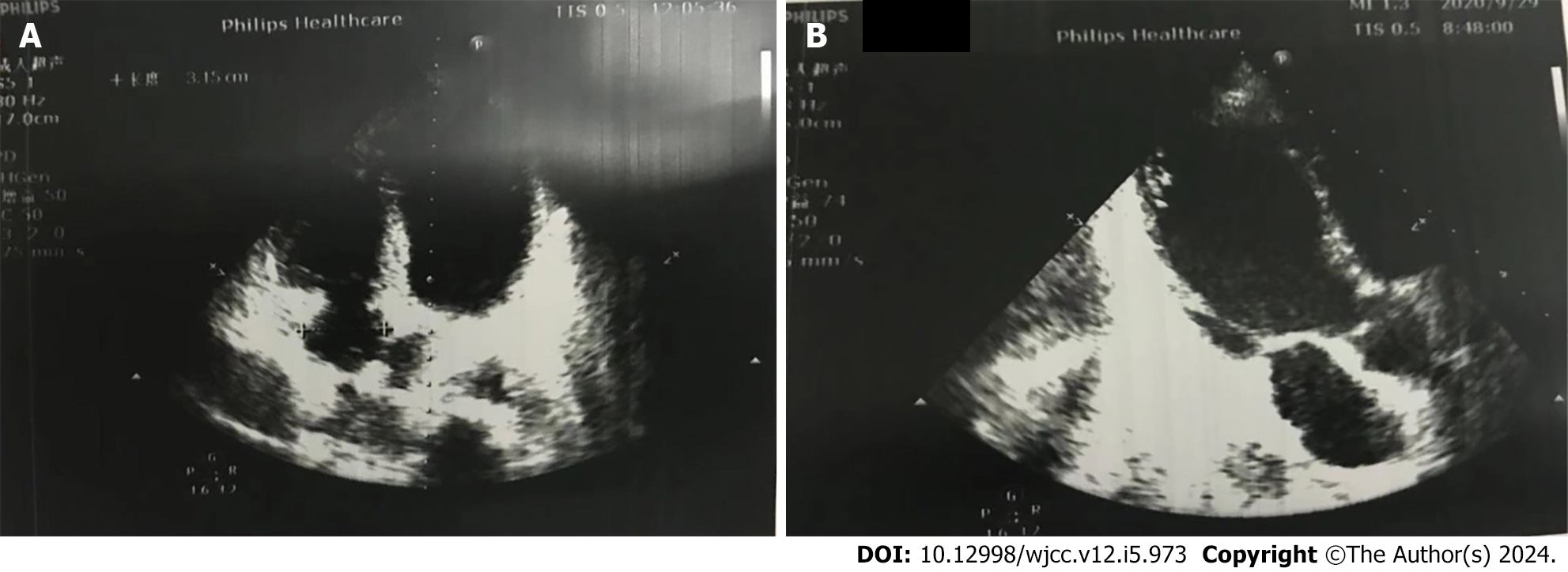
Figure 2 The thrombus sound shadow in the heart was not obvious on the apical four-chamber heart section and the long axis of the left ventricle section.
A: Apical four-chamber heart suggests no thrombus; B: The thrombus was not detected on the long axis of the left ventricle section.
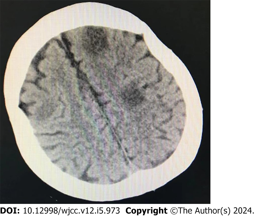
Figure 3 Computed tomography revealed multiple low-density lesions in both hemispheres of the brain, which were cerebral emboli.
- Citation: Bai YB, Zhao F, Wu ZH, Shi GN, Jiang N. Left ventricular thrombosis caused cerebral embolism during venoarterial extracorporeal membrane oxygenation support: A case report. World J Clin Cases 2024; 12(5): 973-979
- URL: https://www.wjgnet.com/2307-8960/full/v12/i5/973.htm
- DOI: https://dx.doi.org/10.12998/wjcc.v12.i5.973


