Copyright
©The Author(s) 2024.
World J Clin Cases. Feb 16, 2024; 12(5): 1025-1028
Published online Feb 16, 2024. doi: 10.12998/wjcc.v12.i5.1025
Published online Feb 16, 2024. doi: 10.12998/wjcc.v12.i5.1025
Figure 1 Urine test of the patient.
A: Before centrifugation; B: After centrifugation (400 g × 5 min); C: Microscopic appearance (40 × 10 times).
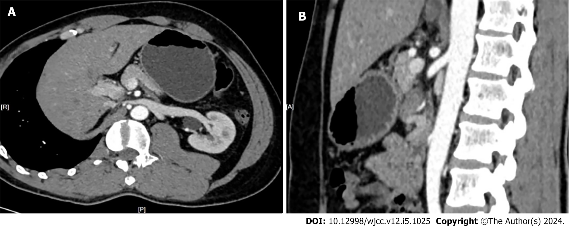
Figure 2 Computed tomography urography of the patient.
A: Transverse plane; B: Sagittal plane.
- Citation: Bai MJ, Yang ST, Liu XK. Hematuria after nocturnal exercise of a man: A case report. World J Clin Cases 2024; 12(5): 1025-1028
- URL: https://www.wjgnet.com/2307-8960/full/v12/i5/1025.htm
- DOI: https://dx.doi.org/10.12998/wjcc.v12.i5.1025


