Copyright
©The Author(s) 2024.
World J Clin Cases. Feb 16, 2024; 12(5): 1018-1024
Published online Feb 16, 2024. doi: 10.12998/wjcc.v12.i5.1018
Published online Feb 16, 2024. doi: 10.12998/wjcc.v12.i5.1018
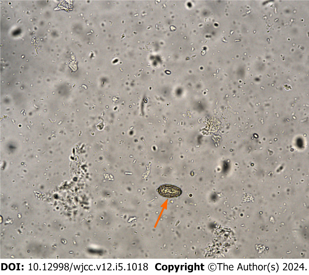
Figure 1 Routine stool examination.
On September 29, 2022, the presence of Clonorchis sinensis eggs (arrow) was confirmed through microscopic examination of stool samples during hospitalization.
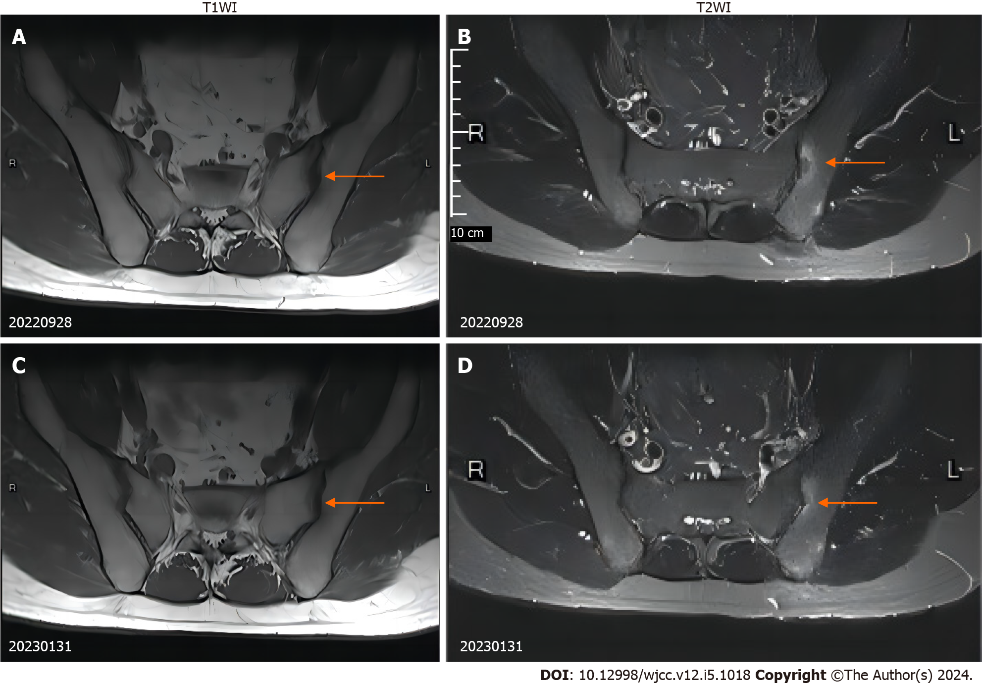
Figure 2 Magnetic resonance imaging of the sacroiliac joint.
A: Axial T1-weighted magnetic resonance imaging (MRI) of the sacroiliac joint showing bone marrow edema in the left sacroiliac joint (September 28, 2022); B: T2-weighted MRI of the sacroiliac joint showing bone marrow edema in the left sacroiliac joint. The patient's condition at the time of admission indicates the presence of inflammation and edema in the left sacroiliac joint (September 28, 2022); C: Axial T1-weighted MRI of the sacroiliac joint showing a reduction in the area around the bone marrow edema in the left sacroiliac joint (January 31, 2023); D: T2-weighted MRI of the sacroiliac joint showing a reduction in the area around the bone marrow edema in the left sacroiliac joint. After treatment, follow-up at 4 months indicated a reduction in inflammation and edema of the sacroiliac joint (January 31, 2023).
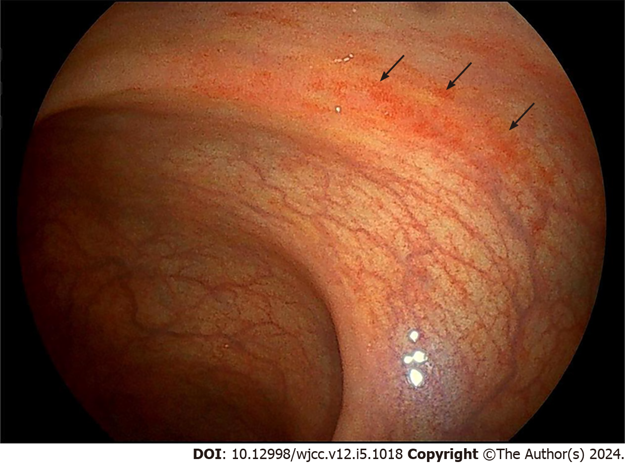
Figure 3 Colonoscopy image.
On September 29, 2022, colonoscopy performed during hospitalization revealed scattered congestion in the descending colon, sigmoid colon, and rectum.
- Citation: Yi TX, Liu W, Leng WF, Wang XC, Luo L. Ankylosing spondylitis coexisting with Clonorchis sinensis infection: A case report. World J Clin Cases 2024; 12(5): 1018-1024
- URL: https://www.wjgnet.com/2307-8960/full/v12/i5/1018.htm
- DOI: https://dx.doi.org/10.12998/wjcc.v12.i5.1018


