Copyright
©The Author(s) 2024.
World J Clin Cases. Feb 6, 2024; 12(4): 859-864
Published online Feb 6, 2024. doi: 10.12998/wjcc.v12.i4.859
Published online Feb 6, 2024. doi: 10.12998/wjcc.v12.i4.859
Figure 1 Upper gastrointestinal imaging.
A: Smooth esophageal wall, large amount of free gas visible in the abdominal cavity, and gas visible around the esophagus; B: Gastric perforation with flow of contrast into the pelvis.
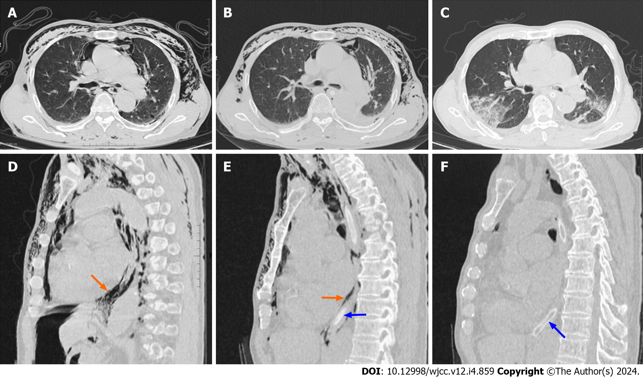
Figure 2 Computed tomography.
A and D: Preoperative computed tomography (CT) showed striated and cast areas without lung texture along the fascial space in the mediastinum and both chest walls. Gas is visible around the esophagus (orange arrow). The blue arrow indicates the gastric tube; B and E: CT on the first postoperative day showed significant reduction of original mediastinal emphysema and chest wall emphysema after surgical treatment; C and F: CT on the sixth postoperative day showed that original mediastinal emphysema and chest wall emphysema largely disappeared.
- Citation: Dai ZC, Gui XW, Yang FH, Zhang HY, Zhang WF. Perforated gastric ulcer causing mediastinal emphysema: A case report. World J Clin Cases 2024; 12(4): 859-864
- URL: https://www.wjgnet.com/2307-8960/full/v12/i4/859.htm
- DOI: https://dx.doi.org/10.12998/wjcc.v12.i4.859


