Copyright
©The Author(s) 2024.
World J Clin Cases. Feb 6, 2024; 12(4): 853-858
Published online Feb 6, 2024. doi: 10.12998/wjcc.v12.i4.853
Published online Feb 6, 2024. doi: 10.12998/wjcc.v12.i4.853
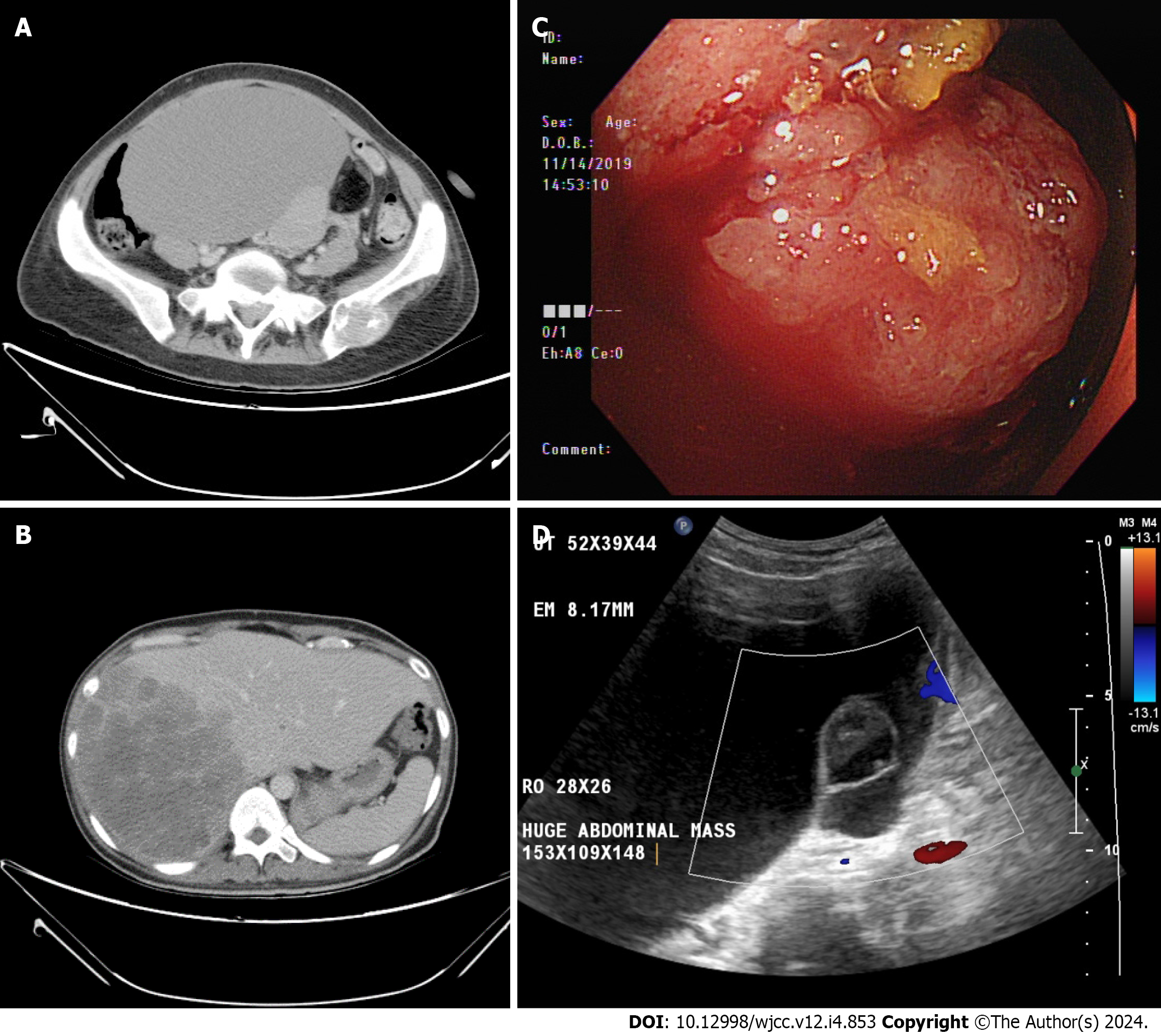
Figure 1 Imaging examinations.
A: The cystic pelvic mass by computed tomography (CT) scan; B: The liver mass by CT scan; C: Colon tumor by colonoscopy; D: The pelvic mass by Doppler trans-abdominal sonography.
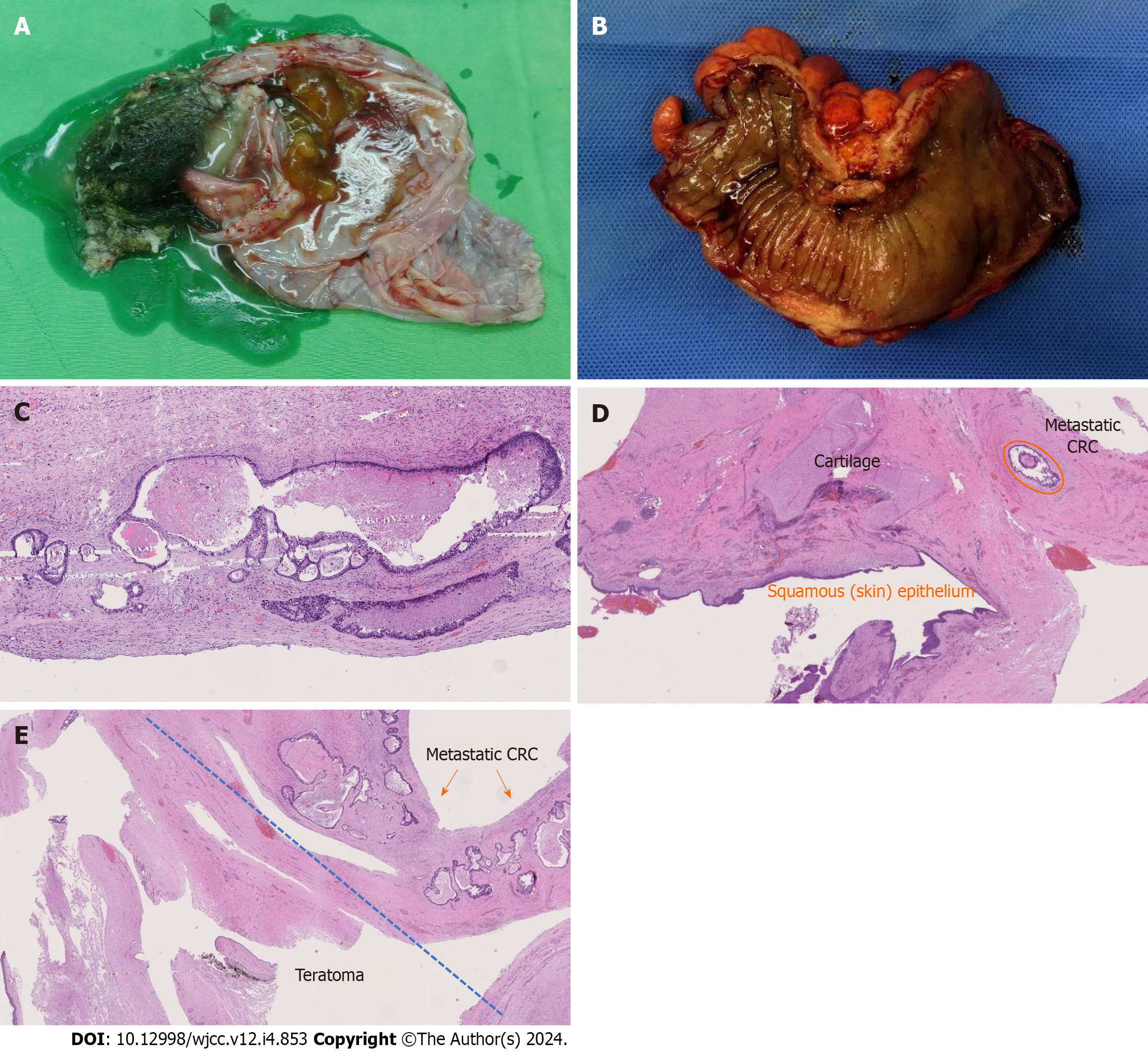
Figure 2 Macroscopic finding and immunohistochemistry test.
A: Macroscopic finding of the ovarian mass; B: Gross view of the primary colon tumor; C: Microscopic view of the primary colon adenocarcinoma; D: Focus of metastatic adenocarcinoma was found in the ovarian tumor; E: Higher magnification of the metastatic colon adenocarcinoma in the mature ovarian teratoma.
- Citation: Wang W, Lin CC, Liang WY, Chang SC, Jiang JK. Adenocarcinoma of sigmoid colon with metastasis to an ovarian mature teratoma: A case report. World J Clin Cases 2024; 12(4): 853-858
- URL: https://www.wjgnet.com/2307-8960/full/v12/i4/853.htm
- DOI: https://dx.doi.org/10.12998/wjcc.v12.i4.853


