Copyright
©The Author(s) 2024.
World J Clin Cases. Feb 6, 2024; 12(4): 847-852
Published online Feb 6, 2024. doi: 10.12998/wjcc.v12.i4.847
Published online Feb 6, 2024. doi: 10.12998/wjcc.v12.i4.847
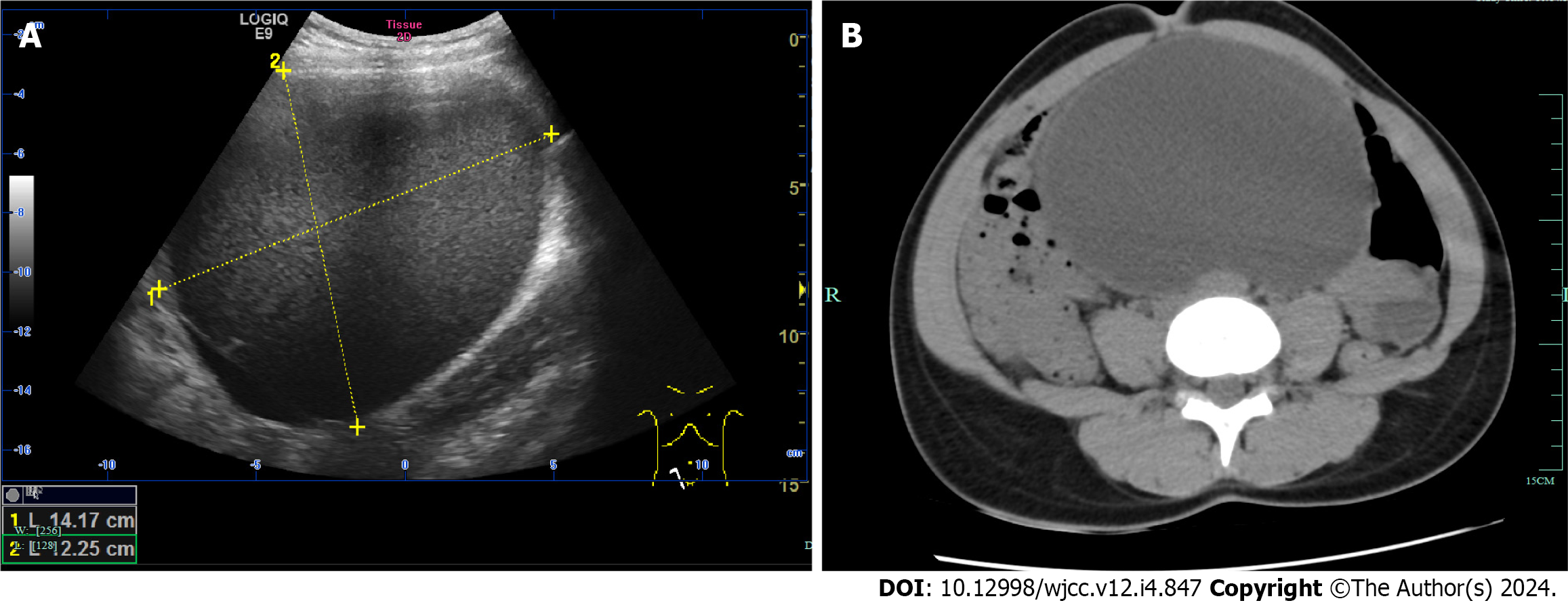
Figure 1 Imaging examinations before laparotomy.
A: Ultrasound image of the patient; B: Abdominopelvic unenhanced computed tomography scan of the patient.
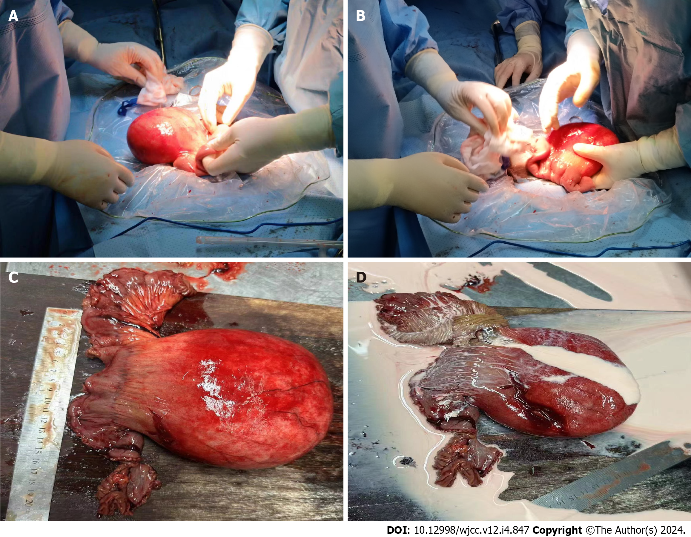
Figure 2 The patient underwent emergency laparotomy and cystectomy.
A: A huge cystic tumor had grown from the jejunum; B: Gastrointestinal surgeons performed a cystectomy; C: The jejunal mesenteric cyst was removed; D: Chylous fluid flowed from the jejunal mesenteric cyst.
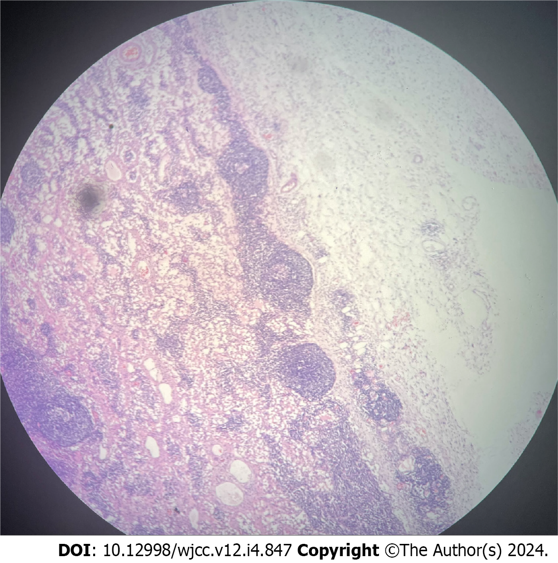
Figure 3 Pathological examination confirmed the diagnosis of cystic lymphangioma of the mesentery.
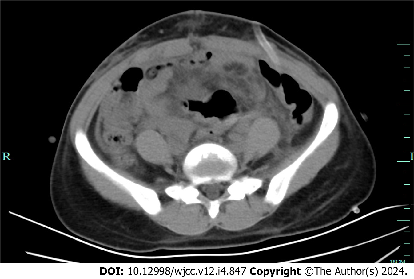
Figure 4 Following the cystectomy, CT showed that the patient’s postoperative recovery was uneventful.
- Citation: Xu J, Lv TF. Rupture of a giant jejunal mesenteric cystic lymphangioma misdiagnosed as ovarian torsion: A case report. World J Clin Cases 2024; 12(4): 847-852
- URL: https://www.wjgnet.com/2307-8960/full/v12/i4/847.htm
- DOI: https://dx.doi.org/10.12998/wjcc.v12.i4.847


