Copyright
©The Author(s) 2024.
World J Clin Cases. Feb 6, 2024; 12(4): 835-841
Published online Feb 6, 2024. doi: 10.12998/wjcc.v12.i4.835
Published online Feb 6, 2024. doi: 10.12998/wjcc.v12.i4.835
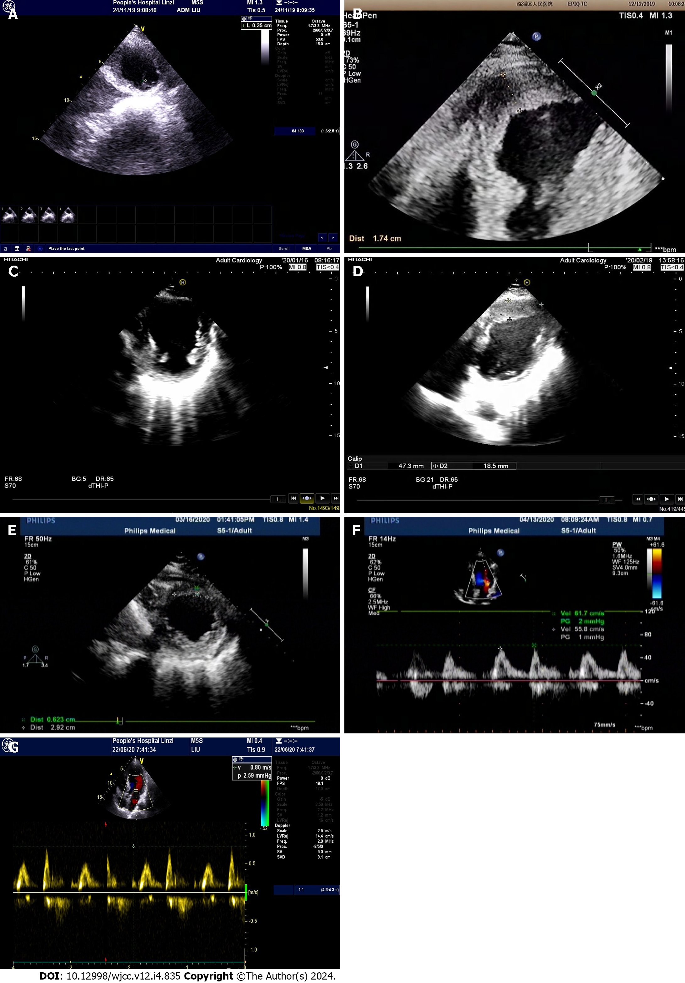
Figure 1 Color ultrasound.
A: Color ultrasound on November 24, showing attached wall thrombosis in the left ventricular apex with a height of 17.6 mm, width of 3.5 mm, and width at the base of 4.8 mm (after enoxaparin was replaced with dabigatran); B: Color ultrasound on December 7, displaying a band-shaped slightly higher echo in the left ventricular apex measuring about 45.3 mm × 42.7 mm × 17.4 mm (13 d after taking dabigatran); C: Color ultrasound on January 16, 2020, revealing the resolution of ventricular thrombus; D: Color ultrasound on February 19, 2020, indicating a left ventricular ejection fraction of 0.45 and an attached wall thrombus measuring about 47.3 mm × 44.7 mm × 18.5 mm; E: Color ultrasound on March 16, 2020, showing an left ventricular ejection fraction of 0.46 and an attached wall thrombus measuring about 32.8 mm × 29.2 mm × 6.2 mm; F: Color ultrasound on April 13, 2020, demonstrating the resolution of the ventricular thrombus; G: Color ultrasound on June 20, 2020, demonstrating the resolution of the ventricular thrombus.
- Citation: Song Y, Li H, Zhang X, Wang L, Xu HY, Lu ZC, Wang XG, Liu B. Individualized anti-thrombotic therapy for acute myocardial infarction complicated with left ventricular thrombus: A case report. World J Clin Cases 2024; 12(4): 835-841
- URL: https://www.wjgnet.com/2307-8960/full/v12/i4/835.htm
- DOI: https://dx.doi.org/10.12998/wjcc.v12.i4.835


