Copyright
©The Author(s) 2024.
World J Clin Cases. Feb 6, 2024; 12(4): 828-834
Published online Feb 6, 2024. doi: 10.12998/wjcc.v12.i4.828
Published online Feb 6, 2024. doi: 10.12998/wjcc.v12.i4.828
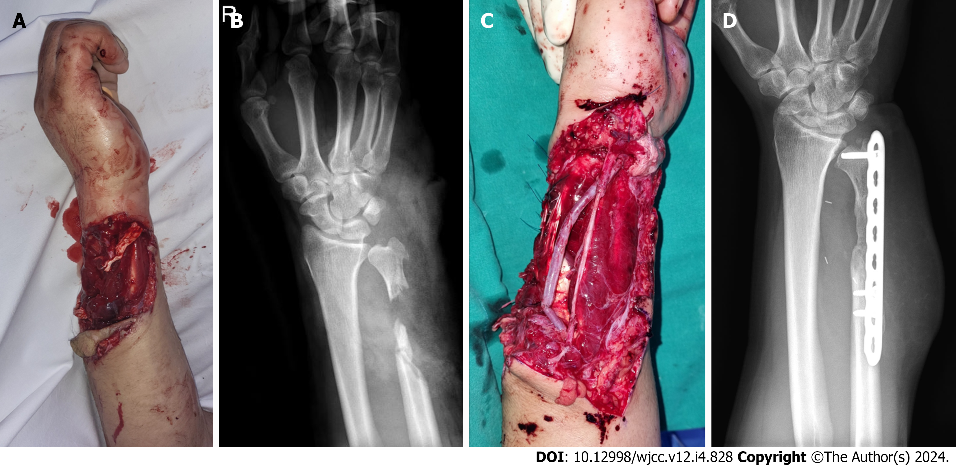
Figure 1 Initial clinical photographs and plain radiographs of the right forearm.
A and B: The 15 cm × 10 cm soft tissue defect of the forearm and open fracture with bone defect of distal ulnar bone were found; C and D: Initially, the ulnar nerve, artery, and tendons were reconstructed, and internal fixation with bone cement were done using the Masquelet technique.
Figure 2 Clinical photographs of the right forearm 10 d after orthopedic management.
A-C: Surgical debridement and negative-pressure wound therapy were performed to prepare the wound bed. Final debridement revealed a 20 cm × 15 cm soft tissue defect with flexor and extensor tendons exposure.
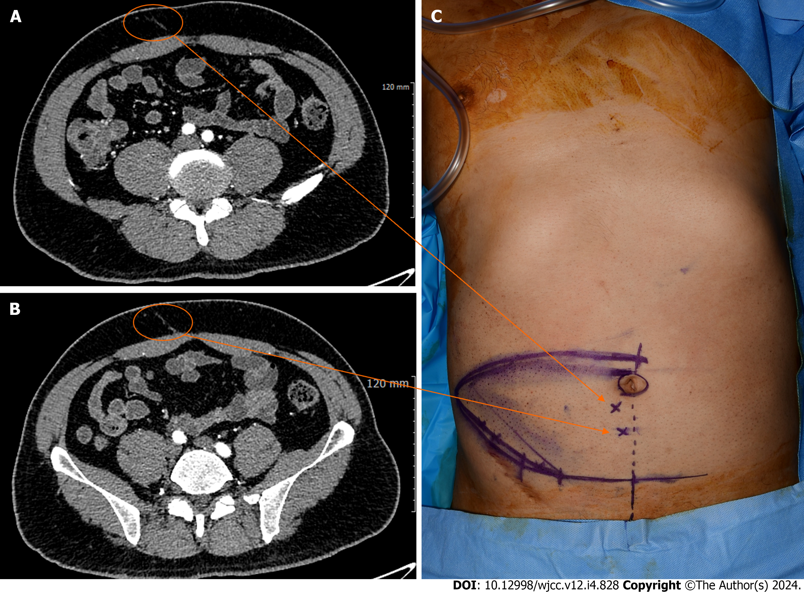
Figure 3 Identification of the deep inferior epigastric artery perforators.
A and B: Preoperative computed tomography findings using 1 mm of slice thickness gave appropriate localization to two deep inferior epigastric artery perforators; C: Intraoperatively, perforators are detected in the paraumbilical area with a handheld Doppler probe, and their positions (X) are marked on the skin.
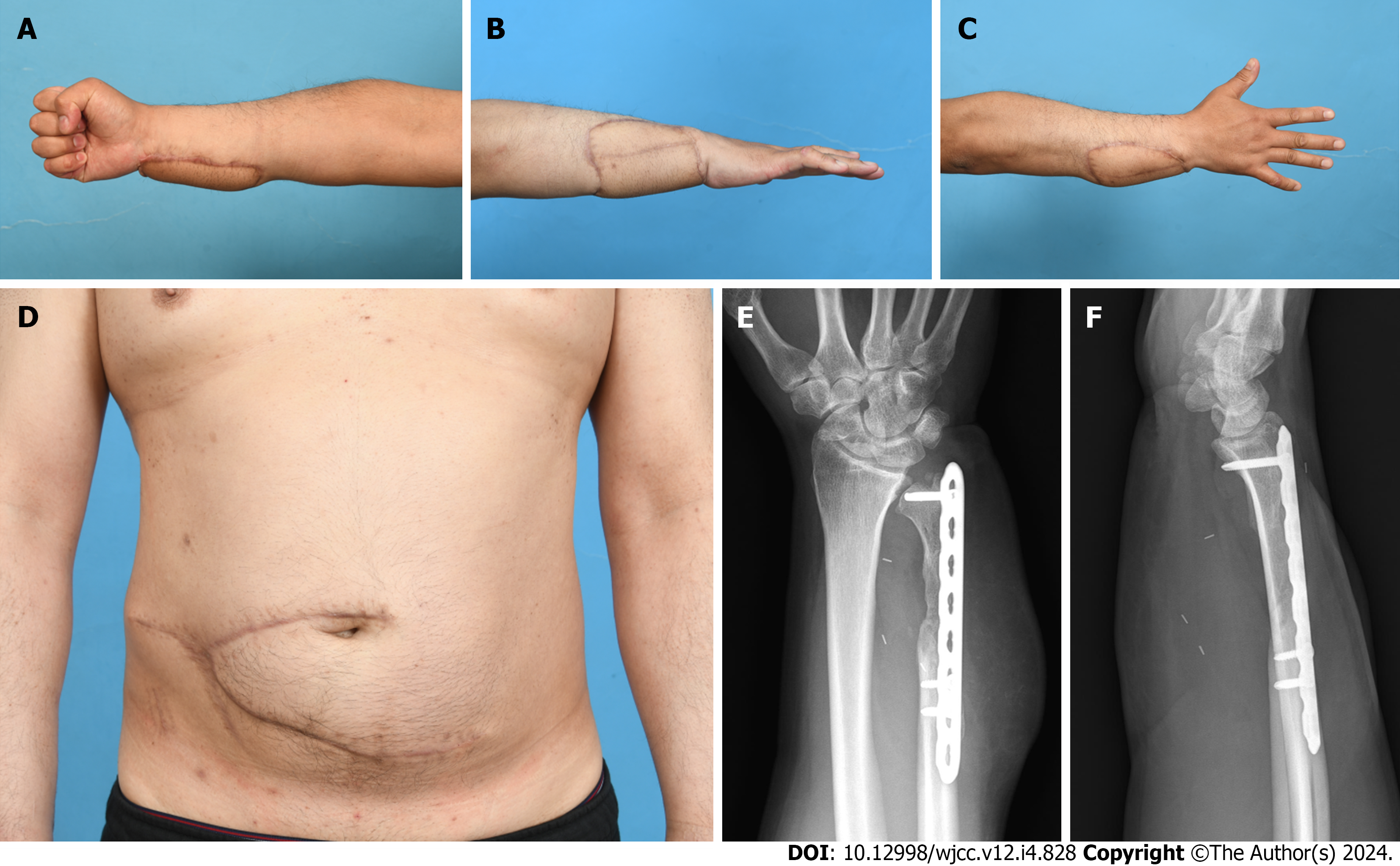
Figure 4 Postoperative clinical photographs and plain radiographs of the right forearm.
A-D: After 6 mo, the pedicled DIEP flap and abdominal donor site were stable without complications; E and F: Ten weeks after the first stage of Masquelet technique, the second stage was performed. Six months later, bone union was achieved.
- Citation: Jeon JH, Kim KW, Jeon HB. Pedicled abdominal flap using deep inferior epigastric artery perforators for forearm reconstruction: A case report. World J Clin Cases 2024; 12(4): 828-834
- URL: https://www.wjgnet.com/2307-8960/full/v12/i4/828.htm
- DOI: https://dx.doi.org/10.12998/wjcc.v12.i4.828


