Copyright
©The Author(s) 2024.
World J Clin Cases. Feb 6, 2024; 12(4): 820-827
Published online Feb 6, 2024. doi: 10.12998/wjcc.v12.i4.820
Published online Feb 6, 2024. doi: 10.12998/wjcc.v12.i4.820
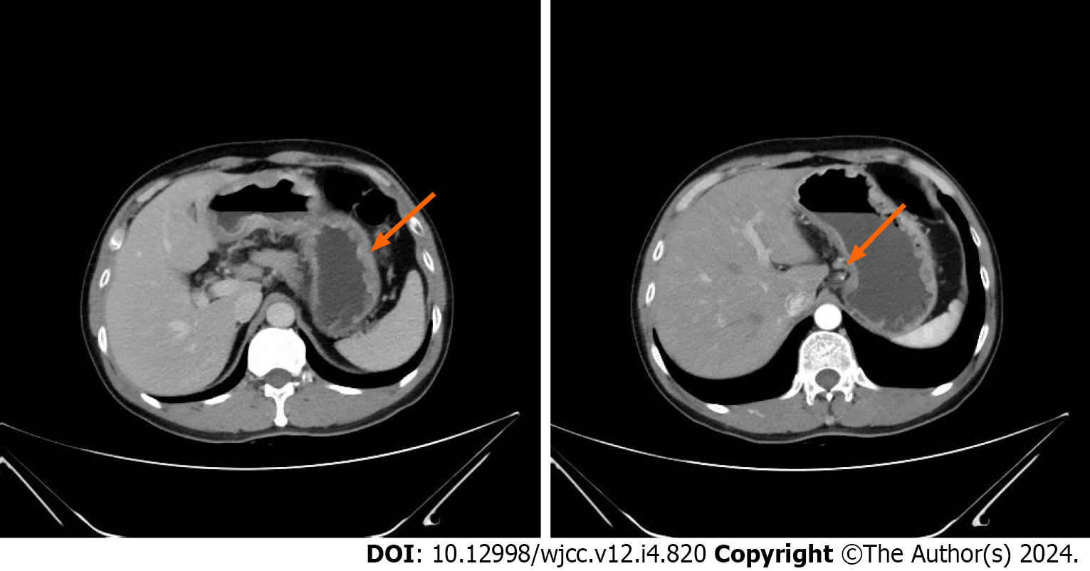
Figure 1 Contrast-enhanced computed tomography images of the whole abdomen, obtained on May 27, 2019.
The computed tomography images revealed abnormal enhancement was observed on the greater curvature of the gastric body and antrum, with enlarged lymph nodes around it (arrows).
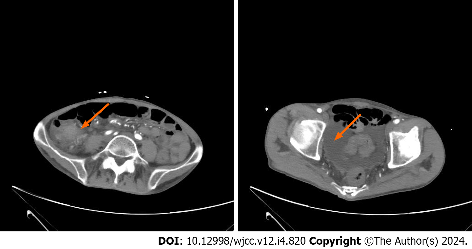
Figure 2 Contrast-enhanced computed tomography images of the whole abdomen, obtained on May 27, 2021.
The computed tomography images revealed significant local thickening and enhancement of the ileum wall, along with abdominal and pelvic effusion and multiple enlarged lymph nodes surrounding the ileum (arrows).
Figure 3 Contrast-enhanced computed tomography images of the whole abdomen after two and four cycles of treatment.
The results of this evaluation indicated stable disease (arrows).
Figure 4 Contrast-enhanced computed tomography images of the whole abdomen after eight, ten, and twelve cycles of treatment.
The results of this evaluation indicated stable disease (arrows).
Figure 5
Variation in cancer antigen 199 levels during second-line treatment.
- Citation: Zhou JH, Yi QJ, Li MY, Xu Y, Dong Q, Wang CY, Liu HY. Inetetamab combined with tegafur as second-line treatment for human epidermal growth factor receptor-2-positive gastric cancer: A case report. World J Clin Cases 2024; 12(4): 820-827
- URL: https://www.wjgnet.com/2307-8960/full/v12/i4/820.htm
- DOI: https://dx.doi.org/10.12998/wjcc.v12.i4.820


