Copyright
©The Author(s) 2024.
World J Clin Cases. Feb 6, 2024; 12(4): 806-813
Published online Feb 6, 2024. doi: 10.12998/wjcc.v12.i4.806
Published online Feb 6, 2024. doi: 10.12998/wjcc.v12.i4.806
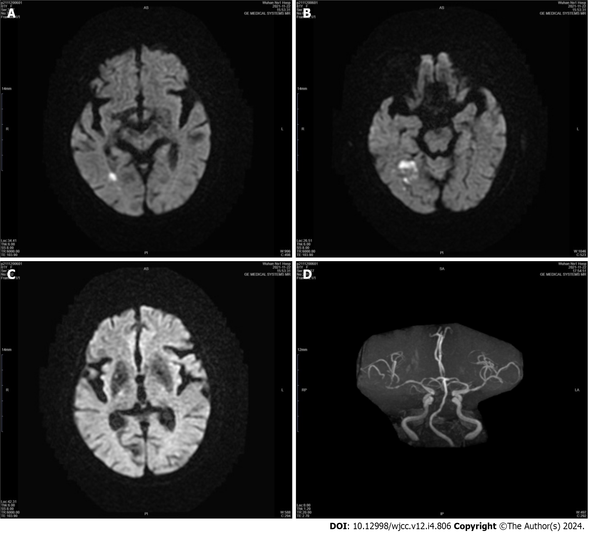
Figure 1 Neuroimaging of Patient 1's first hospitalization.
A-C: Diffusion-weighted imaging showing restricted diffusion in the right temporal lobe, and the basal ganglia, thalamus, and subthalamic nucleus were entirely spared; D: Magnetic resonance angiography of the head showed significant stenosis of the right middle cerebral artery (MCA) and mild stenosis of the left MCA.
Figure 2 Neuroimaging of patient 1’s second hospitalization.
A: Computed tomography perfusion (CTP) showed marked hypoperfusion in the right middle cerebral artery (MCA) territory; B: Cerebral angiography revealed severe stenosis in the M1 segment of the right MCA; C: After successful stenting, CTP showed improved perfusion in the right cerebral hemisphere.
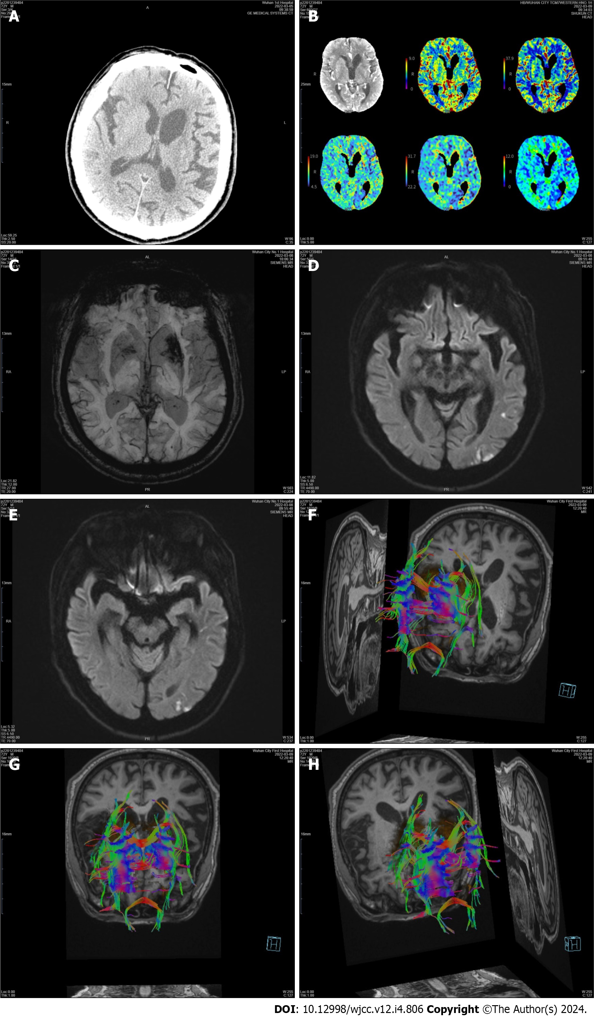
Figure 3 Neuroimaging of patient 2.
A: Computed tomography of the head showed no hyperdensity; B: Computed tomography perfusion showed no abnormalities, with normal perfusion of the basal ganglia; C: Susceptibility weighted imaging showed low signal intensity in the left basal ganglia; D and E: Diffusion-weighted imaging showed an acute patchy ischemic stroke in the left temporal lobe and that the basal ganglia was spared; F-H: Diffusion tensor imaging confirmed fewer white matter fiber tracts on the left side than on the opposite side.
- Citation: Wang XD, Li X, Pan CL. Hemichorea in patients with temporal lobe infarcts: Two case reports. World J Clin Cases 2024; 12(4): 806-813
- URL: https://www.wjgnet.com/2307-8960/full/v12/i4/806.htm
- DOI: https://dx.doi.org/10.12998/wjcc.v12.i4.806


