Copyright
©The Author(s) 2024.
World J Clin Cases. Feb 6, 2024; 12(4): 782-786
Published online Feb 6, 2024. doi: 10.12998/wjcc.v12.i4.782
Published online Feb 6, 2024. doi: 10.12998/wjcc.v12.i4.782
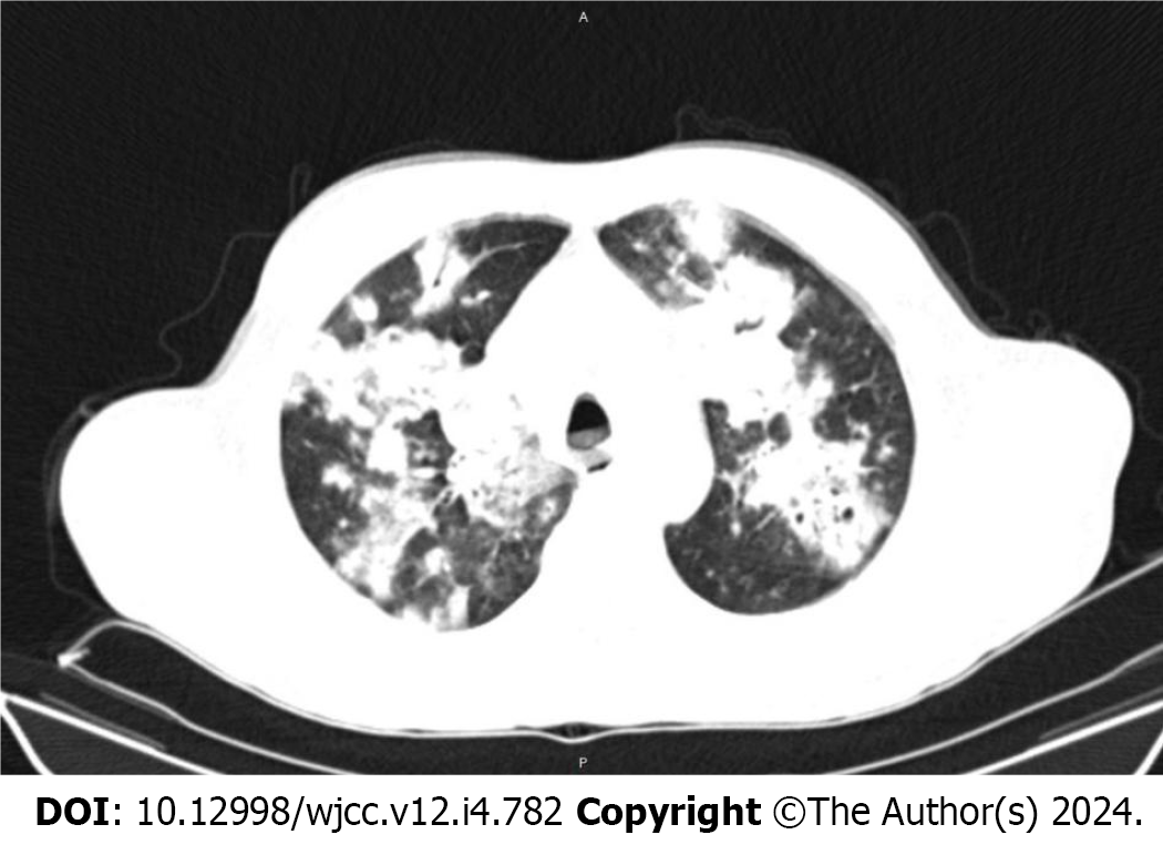
Figure 1 Chest computed tomography of the patient on admission day.
Figure 2 Computed tomography images of the lung.
A: The extracorporeal membrane oxygenation catheter is in the mediastinum; B: Right internal jugular vein; C: Right internal carotid artery.
Figure 3 Computed tomography reconstruction showing that the catheter was only 2 cm in the superior vena cava.
- Citation: Song XQ, Jiang YL, Zou XB, Chen SC, Qu AJ, Guo LL. Accidental placement of venous return catheter in the superior vena cava during venovenous extracorporeal membrane oxygenation for severe pneumonia: A case report. World J Clin Cases 2024; 12(4): 782-786
- URL: https://www.wjgnet.com/2307-8960/full/v12/i4/782.htm
- DOI: https://dx.doi.org/10.12998/wjcc.v12.i4.782


