Copyright
©The Author(s) 2024.
World J Clin Cases. Feb 6, 2024; 12(4): 746-757
Published online Feb 6, 2024. doi: 10.12998/wjcc.v12.i4.746
Published online Feb 6, 2024. doi: 10.12998/wjcc.v12.i4.746
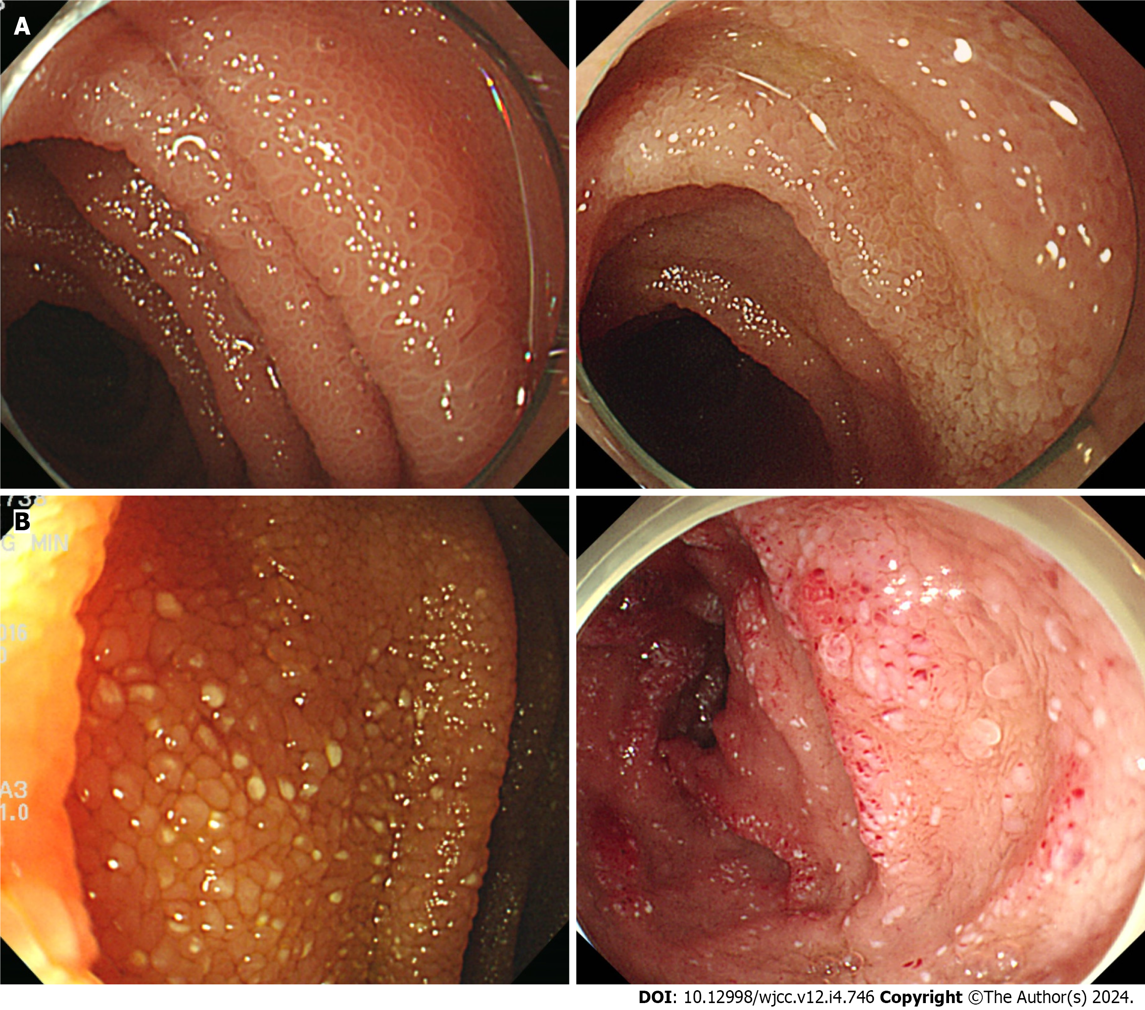
Figure 1 Endoscopic image of the small intestine.
A: Edematous or whitish delineated villi; B: A snowflake appearance.
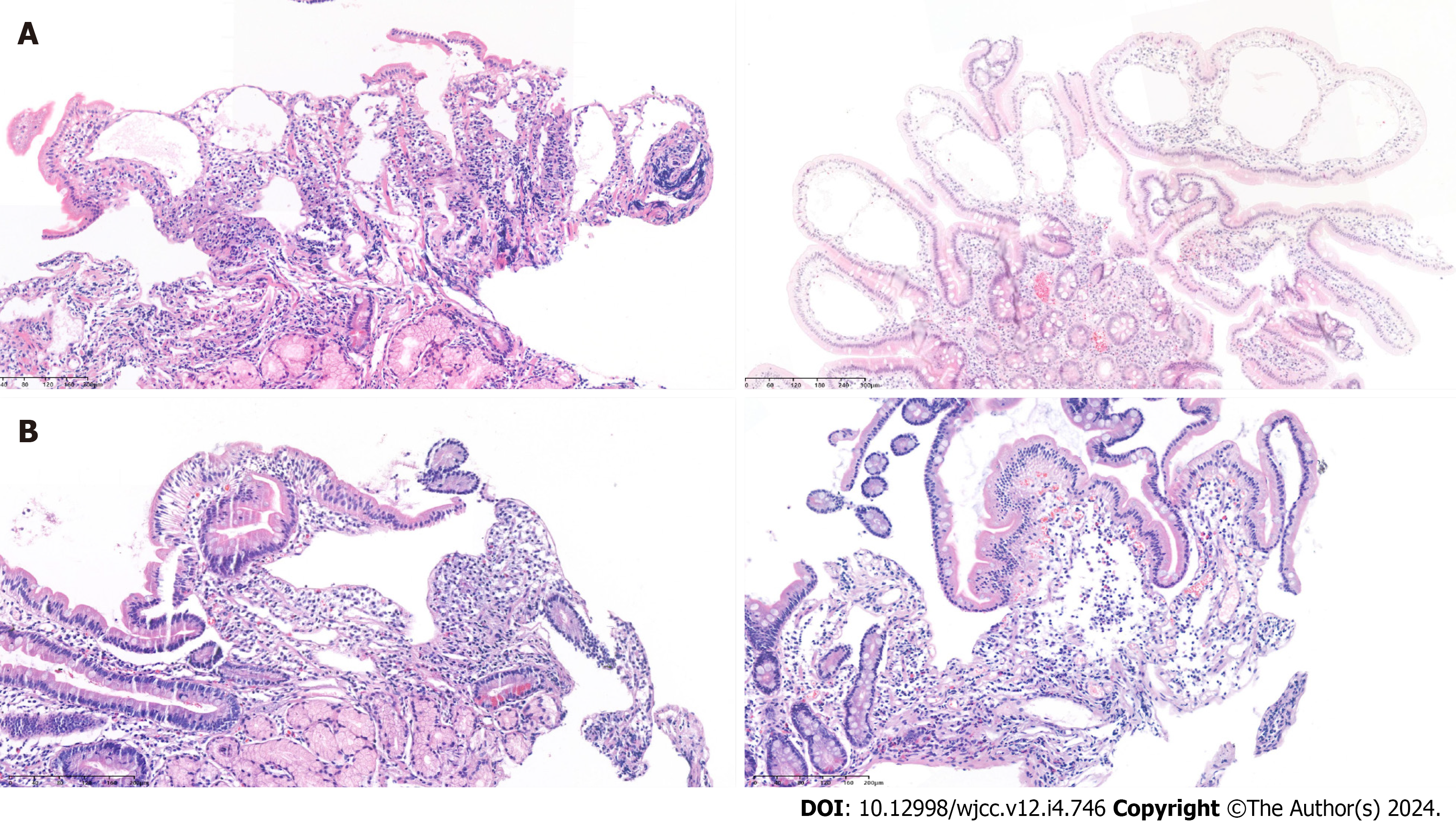
Figure 2 Pathological image of lymphangiectasia in mucosa and submucosa.
A: Marked dilatated lymphatics in the tips of villi; B: Lymphangioma involving mucosa and submucosa.
- Citation: Na JE, Kim JE, Park S, Kim ER, Hong SN, Kim YH, Chang DK. Experience of primary intestinal lymphangiectasia in adults: Twelve case series from a tertiary referral hospital. World J Clin Cases 2024; 12(4): 746-757
- URL: https://www.wjgnet.com/2307-8960/full/v12/i4/746.htm
- DOI: https://dx.doi.org/10.12998/wjcc.v12.i4.746


