Copyright
©The Author(s) 2024.
World J Clin Cases. Dec 26, 2024; 12(36): 6926-6934
Published online Dec 26, 2024. doi: 10.12998/wjcc.v12.i36.6926
Published online Dec 26, 2024. doi: 10.12998/wjcc.v12.i36.6926
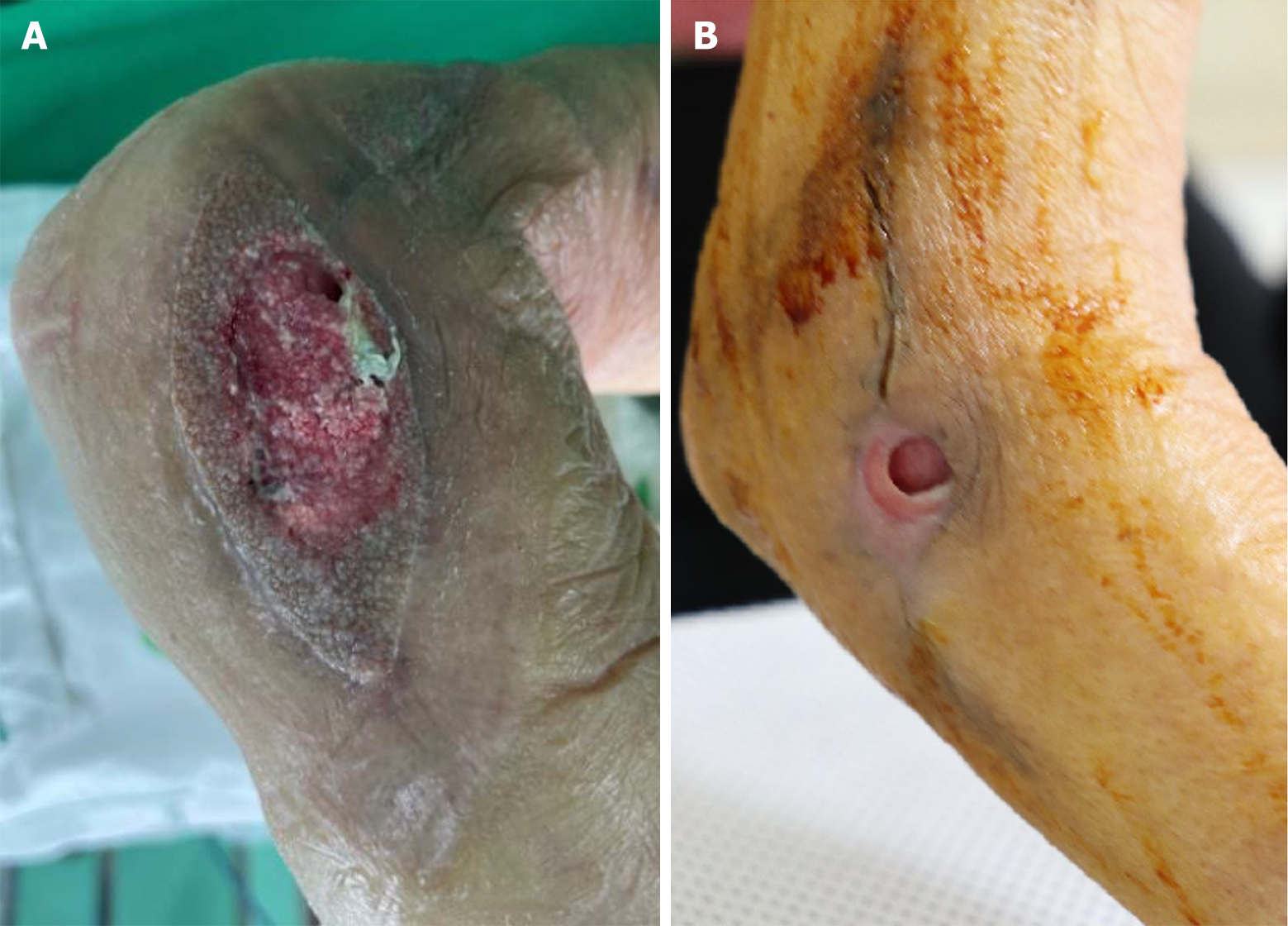
Figure 1 Clinical photographs of the right elbow joint.
A: At treatment onset, the defect (approximately 5.0 cm × 3.0 cm) showed unhealthy granulation tissues, including a partially necrotic change with bone exposure; B: After 4 months of treatment, the dimension of the wound and the amount of exudate were gradually reduced, but the wound stalled without further improvement.
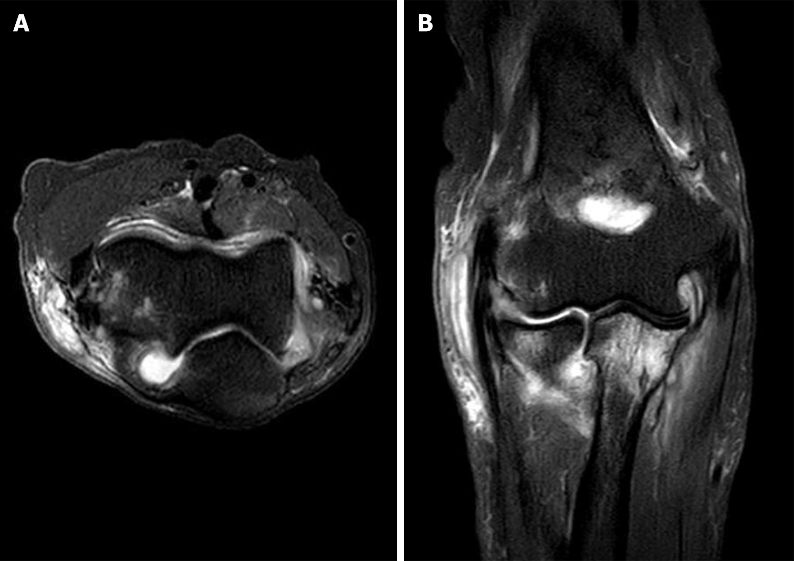
Figure 2 T2-weighted magnetic resonance imaging of the right elbow joint.
Magnetic resonance imaging showing synovial thickening with effusion in the right elbow joint and subcutaneous cystic lesions with adjacent subcutaneous edema at the lateral aspect of the right elbow joint and posterior aspect of the right proximal forearm. A: Axial view; B: Coronal view.
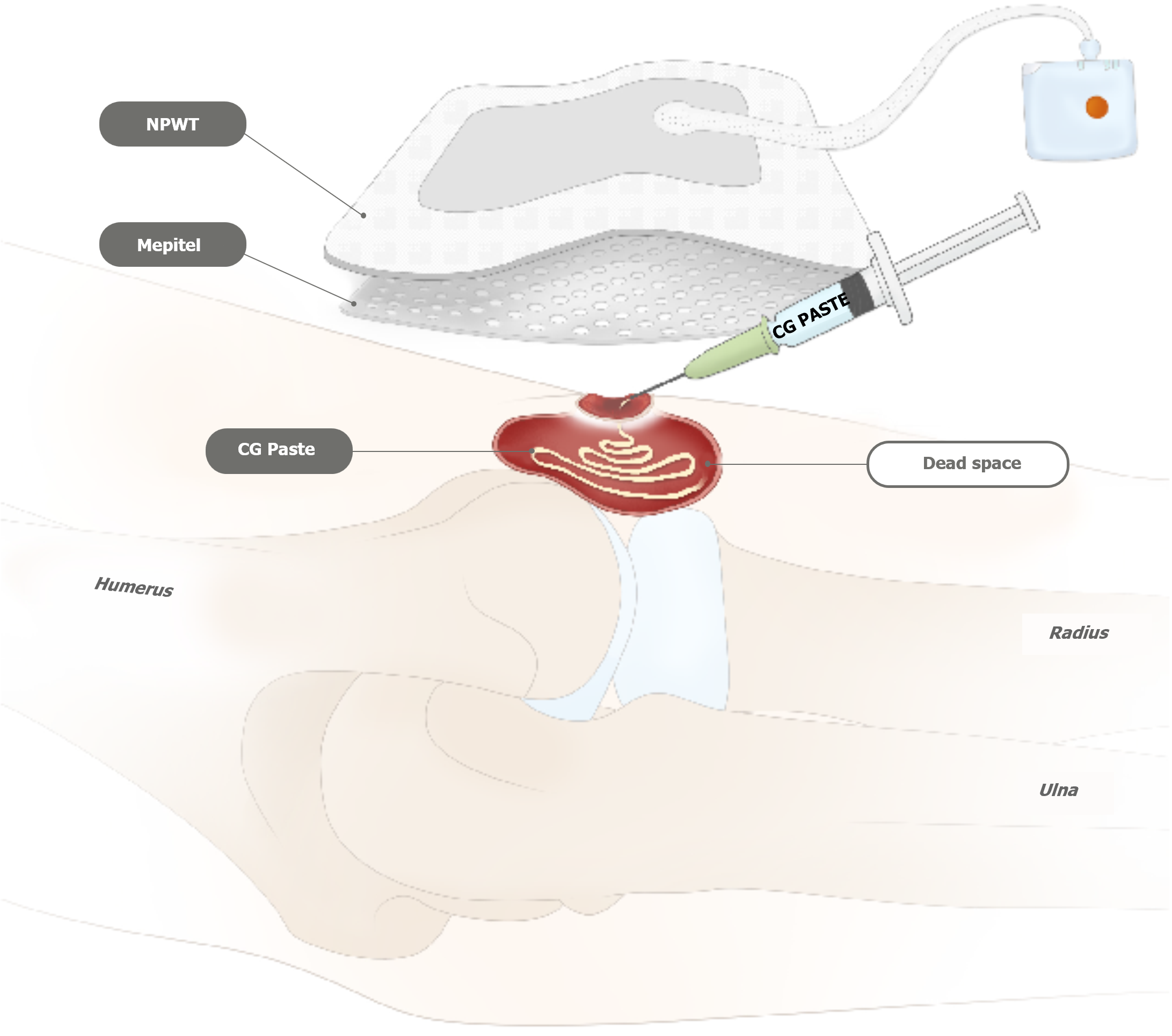
Figure 3 Depiction of injectable paste-type acellular dermal matrix applied in the right elbow joint cavity.
Subsequently, the wound showed gradual reduction in both the dimensions of the raw surface and the amount of exudate. NPWT: Negative pressure wound therapy; CG: Cell and growth factor biotechnology (trade name).
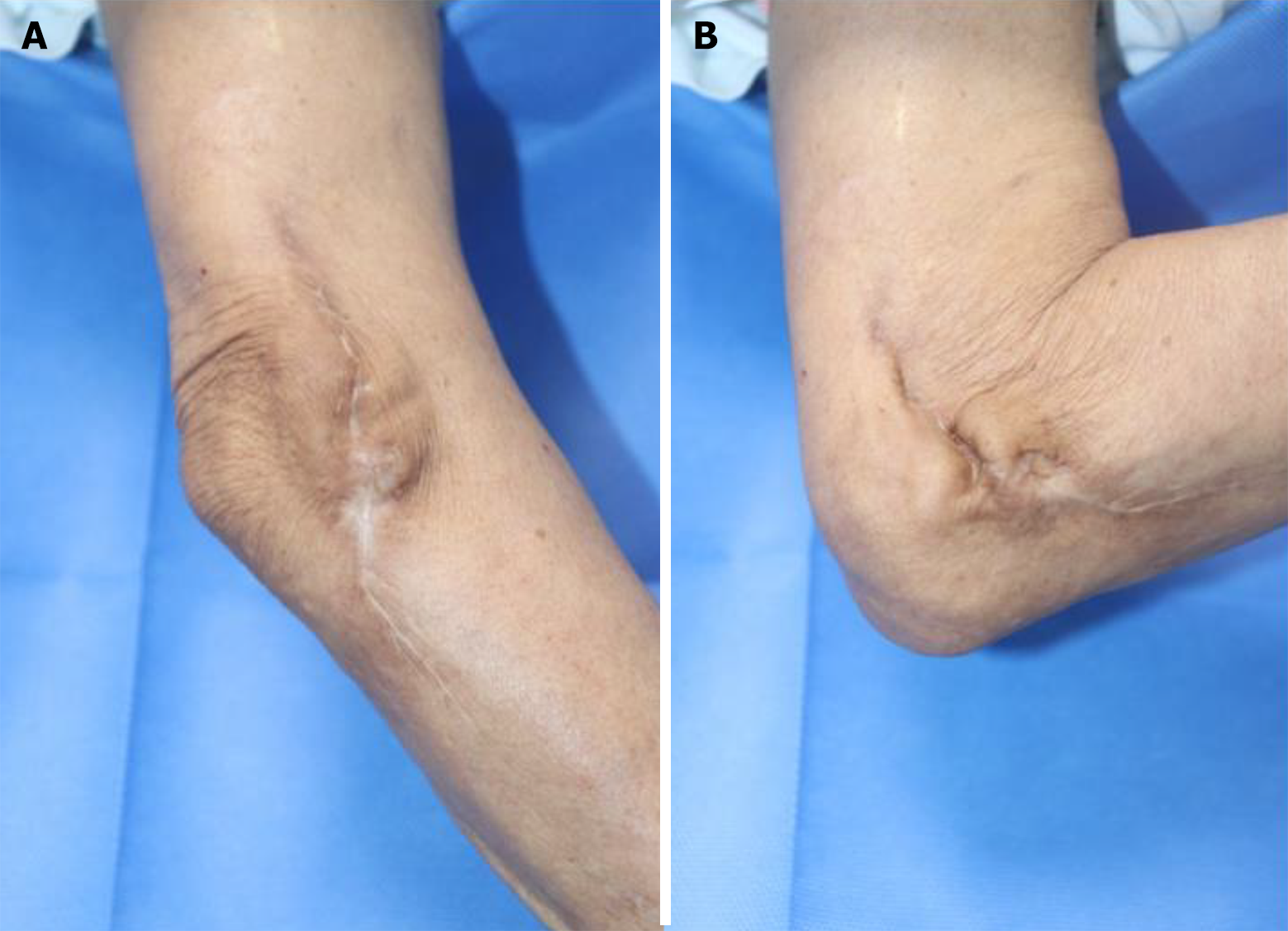
Figure 4 Ten months after complete wound healing, the elbow joint maintains an intact range of motion with no signs of recurrence in the affected area.
A: Elbow extension view; B: Elbow flexion view.
- Citation: Kim JH, Koh IC, Lim SY, Kang SH, Kim H. Chronic intractable nontuberculous mycobacterial-infected wound after acupuncture therapy in the elbow joint: A case report. World J Clin Cases 2024; 12(36): 6926-6934
- URL: https://www.wjgnet.com/2307-8960/full/v12/i36/6926.htm
- DOI: https://dx.doi.org/10.12998/wjcc.v12.i36.6926


