Copyright
©The Author(s) 2024.
World J Clin Cases. Jan 26, 2024; 12(3): 643-649
Published online Jan 26, 2024. doi: 10.12998/wjcc.v12.i3.643
Published online Jan 26, 2024. doi: 10.12998/wjcc.v12.i3.643
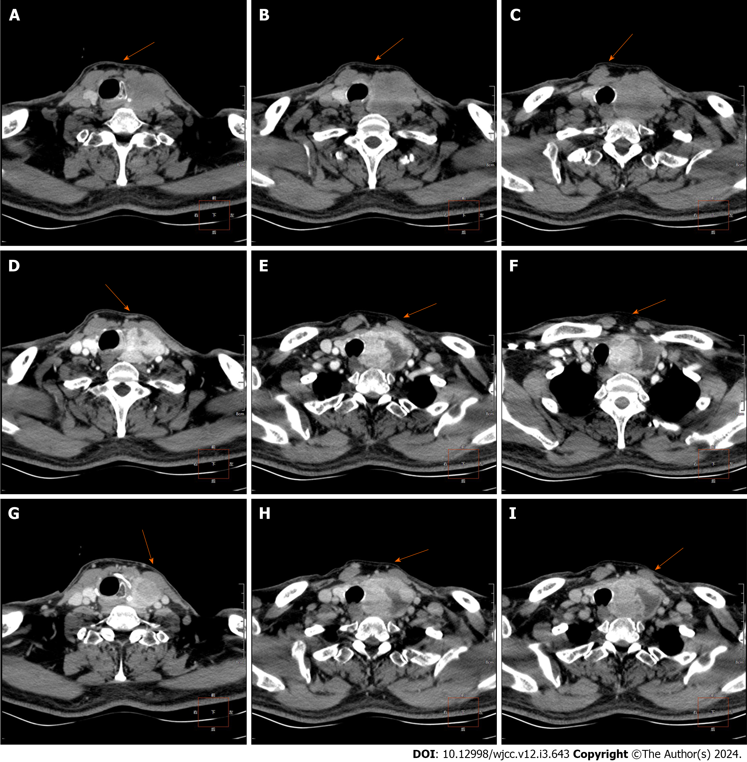
Figure 1 Computed tomography examination before surgery.
A-C: Preoperative computed tomography (CT) scan; D-F: Preoperative enhanced CT (arterial phase); G-I: Preoperative enhanced CT (venous phase).
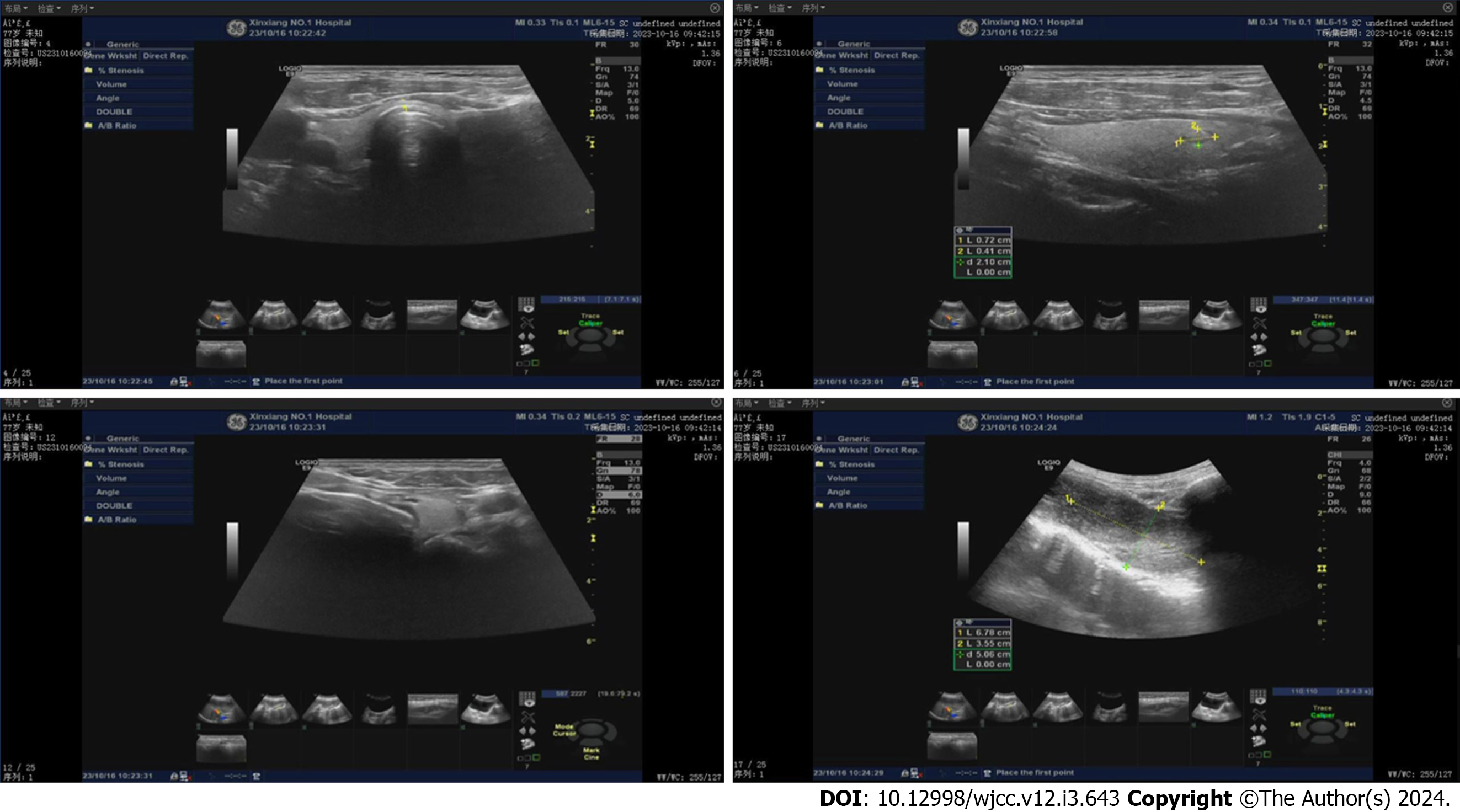
Figure 2 Thyroid and elastic real-time imaging ultrasound.
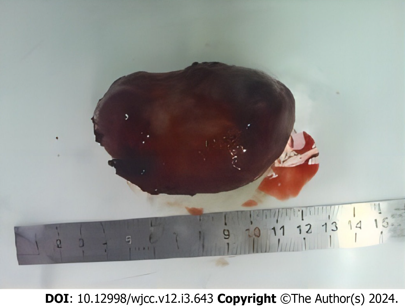
Figure 3 Photos were taken of the size of the left thyroid gland tumor after operation.
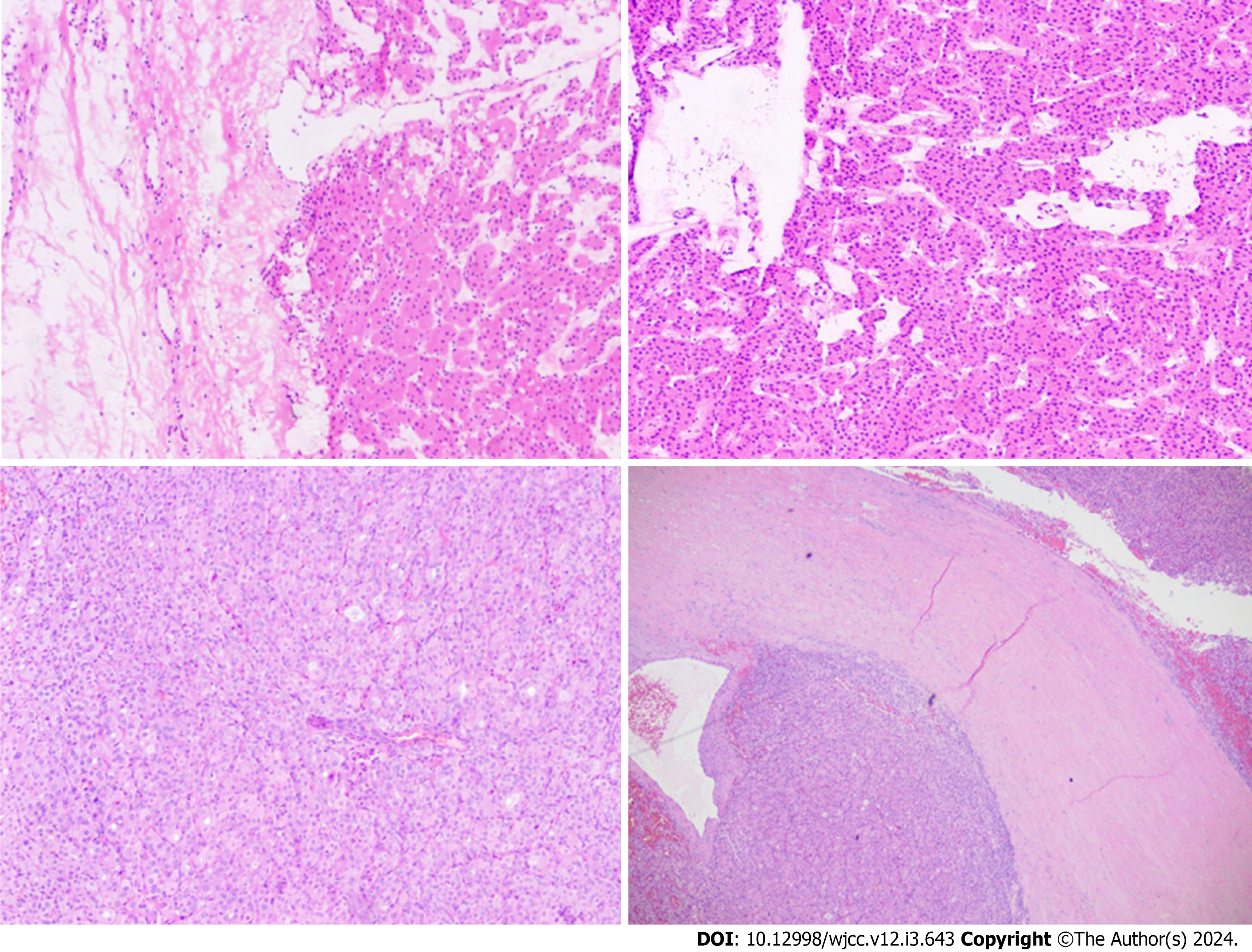
Figure 4 Results of HE staining of the left thyroid gland mass after surgery.
- Citation: Meng YC, Wu LS, Li N, Li HW, Zhao J, Yan J, Li XQ, Li P, Wei JQ. Pathological diagnosis and immunohistochemical analysis of giant retrosternal goiter in the elderly: A case report. World J Clin Cases 2024; 12(3): 643-649
- URL: https://www.wjgnet.com/2307-8960/full/v12/i3/643.htm
- DOI: https://dx.doi.org/10.12998/wjcc.v12.i3.643


