Copyright
©The Author(s) 2024.
World J Clin Cases. Jan 26, 2024; 12(3): 630-636
Published online Jan 26, 2024. doi: 10.12998/wjcc.v12.i3.630
Published online Jan 26, 2024. doi: 10.12998/wjcc.v12.i3.630
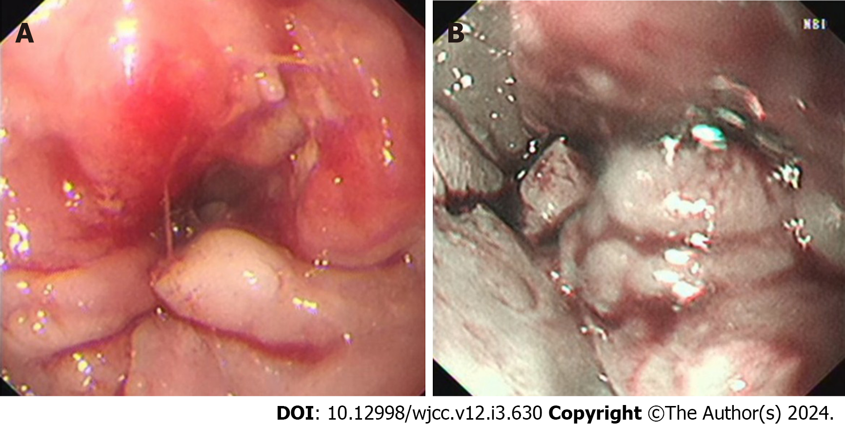
Figure 1 Endoscopic findings and narrow band imaging.
A: Endoscopy; B: Narrow band image.
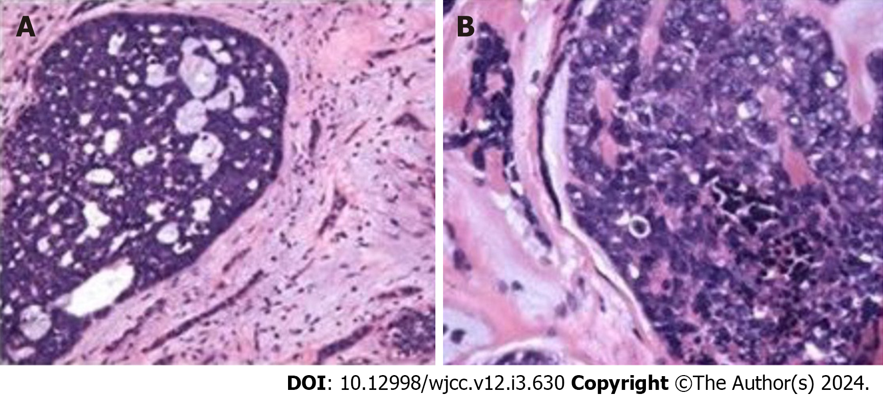
Figure 2 Microscopic appearance of tumor cells.
A: Low power lens; B: High power lens.
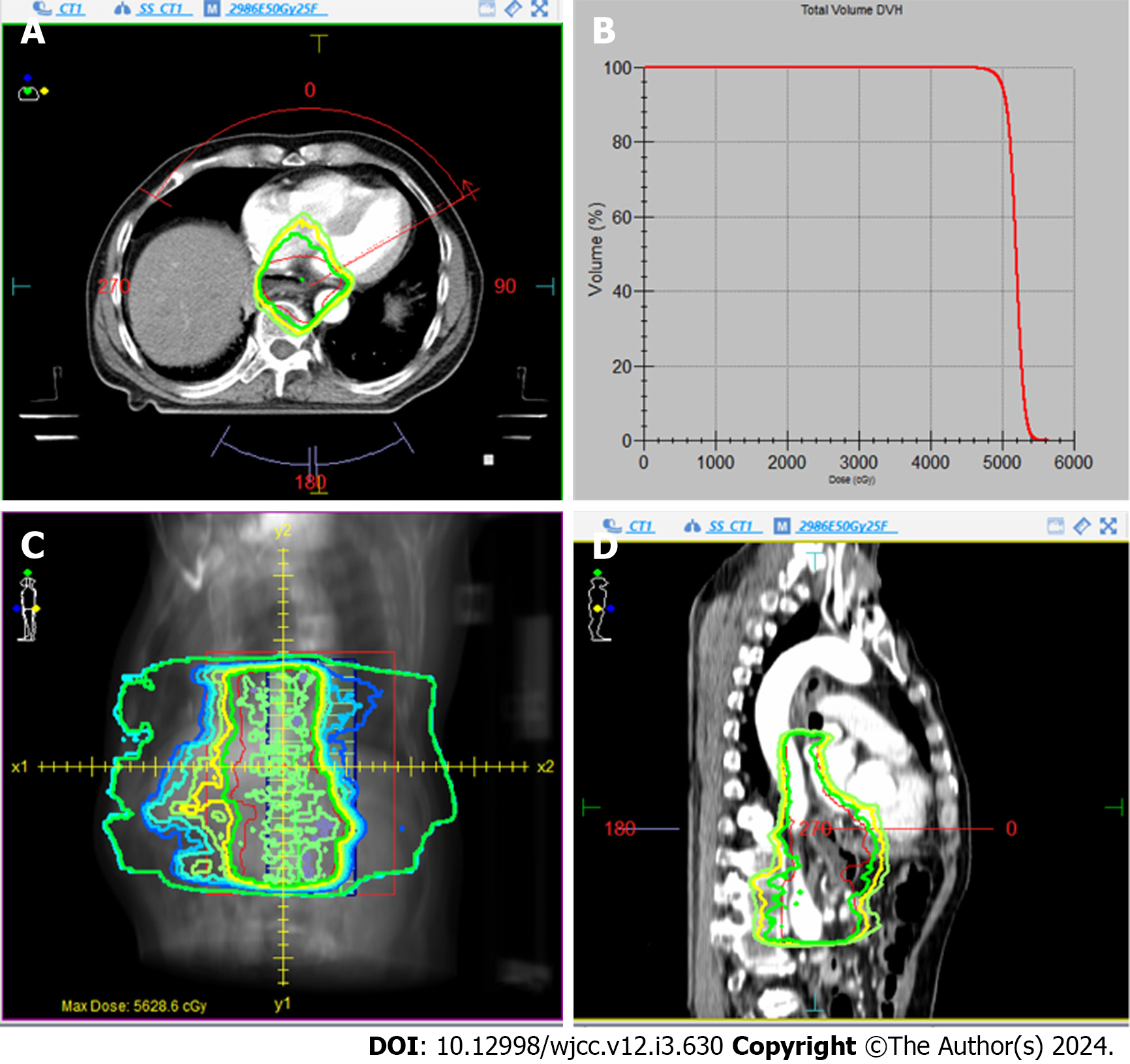
Figure 3 Measurement distribution of radiotherapy targets in the patient.
A-D: Radiotherapy plan (A, C and D) and dose curve histogram (B) diagram.
- Citation: Geng LD, Li J, Yuan L, Du XB. Rare esophageal carcinoma-primary adenoid cystic carcinoma of the esophagus: A case report. World J Clin Cases 2024; 12(3): 630-636
- URL: https://www.wjgnet.com/2307-8960/full/v12/i3/630.htm
- DOI: https://dx.doi.org/10.12998/wjcc.v12.i3.630


