Copyright
©The Author(s) 2024.
World J Clin Cases. Oct 6, 2024; 12(28): 6230-6236
Published online Oct 6, 2024. doi: 10.12998/wjcc.v12.i28.6230
Published online Oct 6, 2024. doi: 10.12998/wjcc.v12.i28.6230
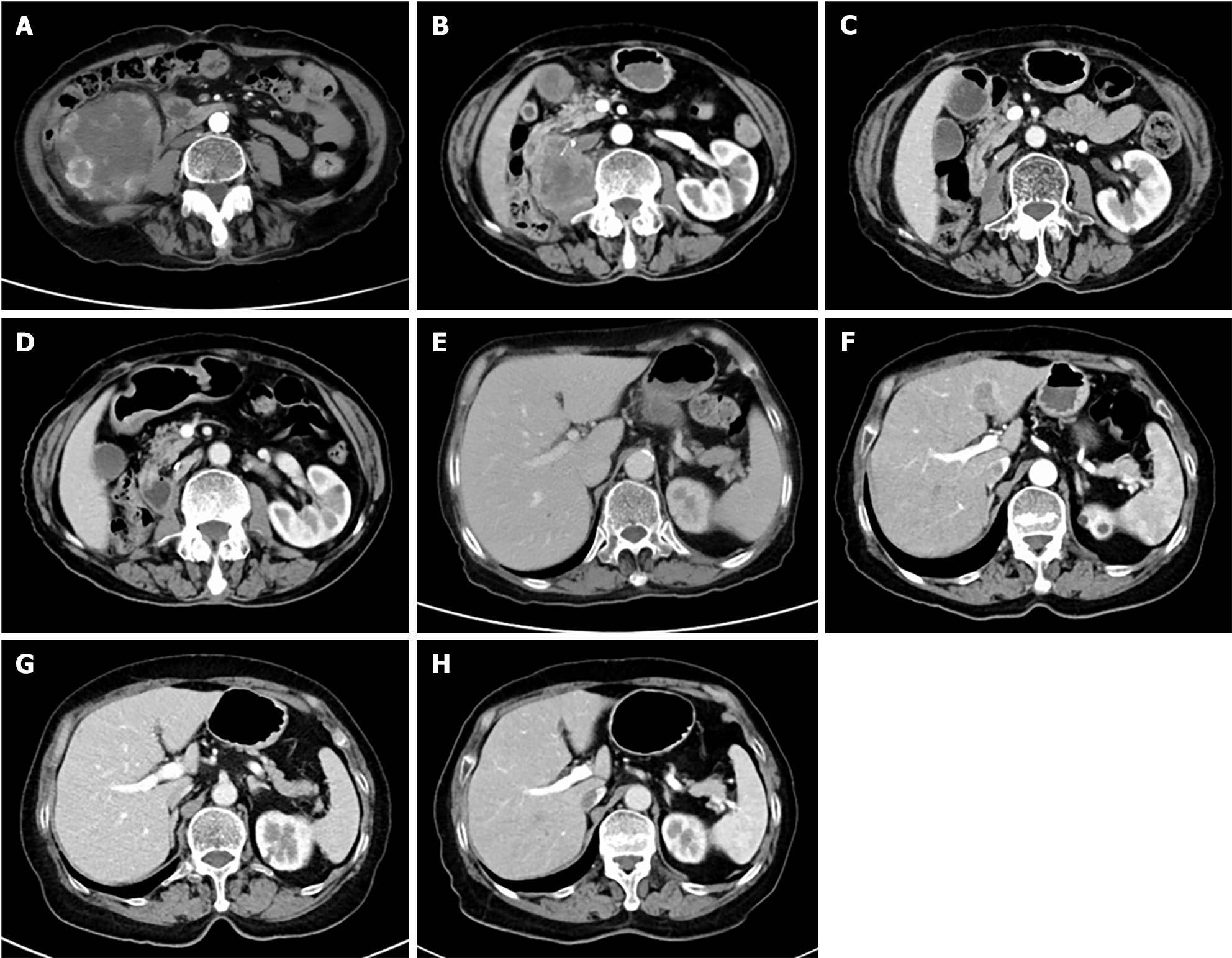
Figure 1 Computed tomography scan findings in the patient with sarcomatoid renal cell carcinoma.
A and E: The computed tomography (CT) scan revealed a tumor in the right kidney, initially believed to be renal cell carcinoma, before surgery in June 2022; B and F: The cancer recurred in the right renal fossa and metastasized to the liver in February 2023; C and G: After six cycles of toripalimab plus pirarubicin chemotherapy, the CT scan showed a notable decrease in the lesions of the right retroperitoneum and liver, indicating a partial response; D and H: After six cycles of toripalimab maintenance therapy, the CT scan showed significant improvement in the original renal tumor area and liver lesions, indicating almost complete remission.
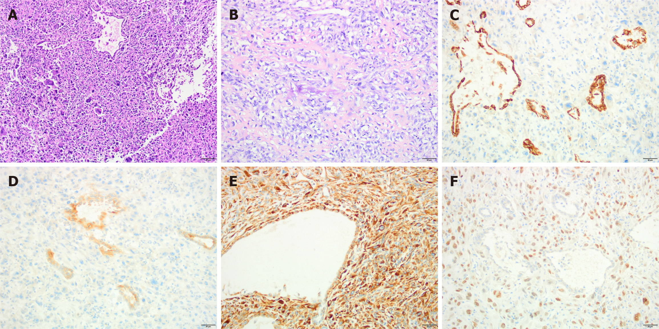
Figure 2 Pathological examinations in the patient with sarcomatoid renal cell carcinoma.
A: 100 ×; B: 200 ×. They displayed a significant proliferation of extremely abnormal spindle cells mixed with tumor giant cells. Additionally, there were areas resembling osteosarcoma and regions with tubular structures. The sarcomatoid component accounts for more than 80% of the tumor; C: The renal cell carcinoma (RCC) component exhibits cytokeratin-pan positivity (400 ×); D: The RCC component was positive for ecological momentary assessment (400 ×); E: Vimentin shows strong immunoreactivity in the sarcomatoid areas and weak immunoreactivity in the renal cell carcinoma areas (400 ×); F: STATB2 exhibits strong immunoreactivity in sarcomatoid areas but not much immunoreactivity in RCC areas (400 ×).
- Citation: Gao MZ, Wang NF, Wang JY, Ma L, Yang YC. Toripalimab in combination with chemotherapy effectively suppresses local recurrence and metastatic sarcomatoid renal cell carcinoma: A case report. World J Clin Cases 2024; 12(28): 6230-6236
- URL: https://www.wjgnet.com/2307-8960/full/v12/i28/6230.htm
- DOI: https://dx.doi.org/10.12998/wjcc.v12.i28.6230


